How PWAS Helped Discover 10 New Alzheimer’s Genes

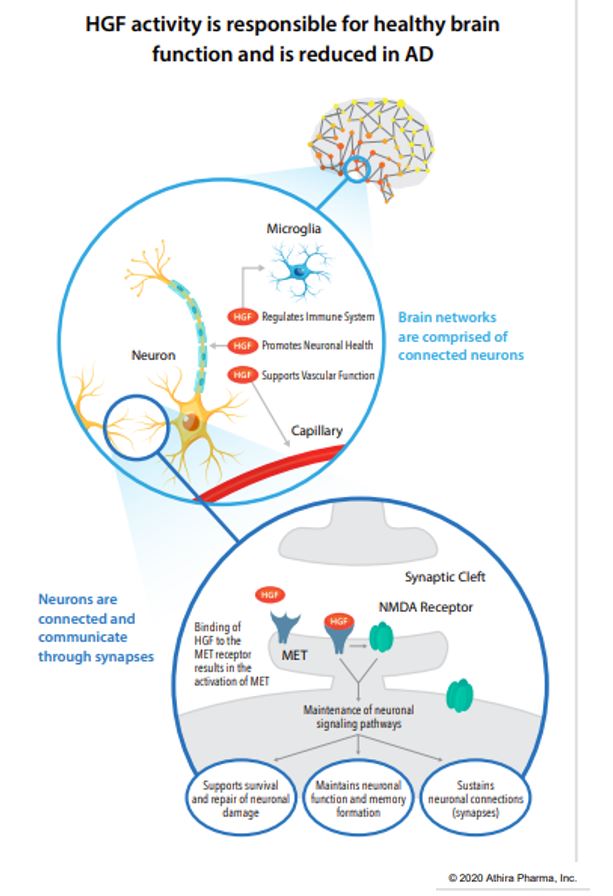
       For quite a while, one of the most common methods of isolating disease-risk-associated genes has been a Genome Wide Association Study (GWAS) which involves genotyping a large number of people and associating various genetic loci, or locations, with the phenotypes, or visible traits, that commonly arise from variance at these loci. However, researchers recently developed an even more informative way of performing these genomic analyses, using it to discover 10 new genes that may modify risk of Alzheimer’s disease (AD)! This new method is very similar to GWAS but adds another layer of data regarding protein function, becoming a Proteome-Wide Association Study (PWAS).

       PWAS expands upon the information provided by GWAS by analyzing the type of mutation present and quantifying the change in production or functionality of the protein produced by that gene in comparison to controls. Next, protein function scores are correlated to the phenotypes of subjects with those genes to confirm the expected effects. In analysis of a binary phenotype (a trait that is either present or absent with no intermediate presentation) a strong correlation is derived when subjects with a disorder have a significantly different functional effect score than controls, confirming PWAS’s prediction that the protein is less (or more) functional in the mutated form.

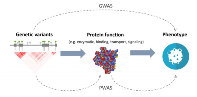
Figure 1. A diagram depicting the difference in analyses between Genome-Wide Association Studies and Proteome-Wide Association Studies.

       The additional information provided by PWAS allows detection of associations that are not detectable by GWAS. Researchers at Emory University recently took advantage of this, using PWAS to discover 10 new genes associated with AD risk. To begin, they isolated 1,475 genes whose abundance is genetically controlled and analyzed their AD-risk score in a GWAS dataset (71,880 cases, 383,378 controls). Of the 1,475 genes, only 13 were related to AD risk in the GWAS dataset.

       After further analyses with PWAS, including causality tests and adjustments for APOE status, 11 genes remained with evidence for causality of AD. Only 1 of these genes, ACE, had previously been strongly correlated with AD but the other 10 are relatively new discoveries! Those genes include syntaxin 4 and 6, DOC2A, SNX32, ICA1L, cathepsin H, CARHSP1, LACTB, RTFDC1, and PLEKHA1. Although little is known about their relationship to AD pathogenesis, researchers will undoubtedly begin researching how these genes relate to AD risk and development. In time you may even see treatments emerging targeting these genes or proteins.

       Even more interestingly, several of these genes impact molecular pathways that are not widely considered part of the disease model for AD, such as LACTB which is a mitochondrial protein (with mitochondrial dysfunction only getting attention as a possible mechanism of AD development within the last few years) and PLEKHA1 which mediates transmembrane signaling more generally. This suggests that there are as-of-yet undiscovered or under-researched factors that relate to AD risk and pathogenesis, opening the door for new treatments and a more complete understanding of the disease itself.

Facebook
Twitter
Email
Sources:
PWAS x GWAS? Proteome Analysis Nets 10 New Alzheimer’s Genes [Internet]. Alzforum. 2021. Available from: https://www.alzforum.org/news/research-news/pwas-x-gwas-proteome-analysis-nets-10-new-alzheimers-genes
Brandes, N., Linial, N, & Linial, M. PWAS: Proteome-Wide Association Study [Internet]. bioRxiv. 2019. Available from: https://www.biorxiv.org/content/10.1101/812289v1.full

Pollution and Plaques: How Your Environment Impacts Neurodegeneration

       It was previously thought that fine particulate matter, produced by car exhaust and wildfires among other things, makes its way into the brain where it damages neurons and glia directly or damages brain vasculature leading to vascular dementia. However, young people in Mexico City (a high pollution area) have also been shown to already possess amyloid plaques, tau neurofibrillary tangles, and alpha-synuclein aggregates in the brain suggesting that pollution may also lead to dementia via non-vascular mechanisms. Two more recent studies, led by Gil Rabinovici at UC San Francisco and Diana Younan at USC Los Angeles, decided to expand on this research.

       Gil and his colleagues used the IDEAS cohort PET scans and related the amyloid burden to air pollution levels in the ZIP codes of participants 1-2 years and 13-15 years before their scan. Those who lived in areas of higher pollution (measured in µg/m3 or PM2.5) were 1.15 times more likely to have a positive amyloid PET per 4 µg/m3 increase. Melinda Power of George Washington University wants to expand this study further to include measures of residential history and occupational pollution exposure, warning that ‘Area-level exposure’, such as that of an entire ZIP code, ‘may or may not be representative of individual-level exposure’.

       Meanwhile, Diana Younan and colleagues looked for a relationship between air pollution and brain atrophy of AD-associated structures in cognitively normal older women. They used MRIs from the WHIMS cohort (712 women who received an MRI scan in 2005-2006 and again in 2010-2011) and their addresses (taken twice a year). They observed that for every 2.82 µg/m3 increase in PM2.5, participants experienced gray matter atrophy associated with a 24% increase in AD risk! What is even more concerning is that these findings, and Gil’s, came from participants who, largely, live in areas that are within the Environmental Protection Agency’s acceptable pollution levels. This means that toxin levels that the EPA determines to be “safe” are already increasing the risk of cognitive impairment, highlighting the need for cleaner/greener ways of living.

       Overall, these studies link pollution to dementia widely and to AD dementia in the form of amyloid accumulation and brain atrophy. The fact that the results remain consistent despite different cohorts and different testing paradigms is compelling but epidemiologists want to see further replication, stating that the direction of the relationship between air pollution and specific neuropathologies is not clear enough yet. Luckily, air quality in the US has improved steadily since 1990 when the Clean Air Act was amended but, as these studies show, these patterns exist even at the currently accepted levels of air quality, leaving room for improvement.

Facebook
Twitter
Email
Sources:
You Are What You Breathe: Polluted Air Tied to Plaques, Brain Atrophy [Internet]. 2020. Alzforum. Available from: https://www.alzforum.org/news/research-news/you-are-what-you-breathe-polluted-air-tied-plaques-brain-atrophy.
Iaccarino, L., et al. Association Between Ambient Air Pollution and Amyloid Positron Emission Tomography Positivity in Older Adults With Cognitive Decline [Internet]. JAMA Neurology. 2020. Available from: https://pubmed.ncbi.nlm.nih.gov/33252608/.
Younan, D., et al. PM2.5 associated with gray matter atrophy reflecting increased Alzheimers risk in older women [Internet]. Neurology. 2020. Available from: https://pubmed.ncbi.nlm.nih.gov/33208540/.

Further Breakthroughs in AD Testing

       A couple months ago we posted a blog about a blood test for Alzheimer’s disease (AD) that changed the game of diagnosing AD. Unsurprisingly, the scientific community immediately began researching and attempting to improve the test and already they have made significant progress! This week we will break down what we have learned about the AD blood test, why it matters, and what remains to be seen.

       Amyloid-β (Aβ) residues and various sub-types of the AD biomarker protein tau are present both in the blood and in the brain. The basis of the AD blood test involves identifying how much of the Aβ and tau sub-types (primarily p-tau181 and p-tau217) are present in serum and correlating these levels to those in the brain to assess increased risk of AD. The initial findings promised a test to identify AD biomarkers, and therefore risk of developing AD, long before symptoms begin.

       Continued research supported the accuracy of the tests ability to identify AD biomarkers years before PET imaging and decades before onset of symptoms. The blood test was recently approved under the Clinical Laboratory Improvements Amendment protocol meaning that it is an accepted method of testing human samples outside of research settings.

       Researchers continued to compare the accuracy of results of p-tau181 levels, Aβ42/40 ratios, and neurofilament light (NfL) testing to see which may be a better predictor of assessing AD risk. P-tau181 levels combined with NfL testing proved the best method. Together, they can differentiate between AD and other dementing illnesses such as Frontotemporal Dementia and Progressive Supranuclear Palsy with 88% accuracy!

       On top of its use for diagnostic purposes, this testing method can also predict disease progression. One study showed that groups with higher p-tau181 levels progressed more quickly over the course of 2.5 years compared to those with lower p-tau181 levels. Other studies suggest this trend may even apply at the individual level with testing of p-tau181 and NfL predicting progression to AD over the course of 4 years in two separate studies of 107 and 74 participants respectively. Once again, the predictions were 88% accurate!

       However, despite all of this promise, some experts in the field aren’t convinced and would like to see more results. Oskar Hansson of Lund University says that he believes tau serum testing is a great method for diagnosing AD in those with MCI/dementia but that diagnosing preclinical AD will require a combination test for multiple blood-based markers. Furthermore, while p-tau181 tests have been quite useful, p-tau217 is seemingly even more useful with one study suggesting it might have twice the predictive power in comparison to p-tau181. As you can see, tau serum blood tests have opened up a new door into the progression of AD but studies remain to be done on how to most effectively use this new tool.

Facebook
Twitter
Email
Source:
Plasma Aβ Test Wins Approval – Are p-Tau Tests Far Behind? [Internet]. 2020. Alzforum. Available from: https://www.alzforum.org/news/research-news/plasma-av-test-wins-approval-are-p-tau-tests-far-behind

Aducanumab: Third Time’s the Charm?

        This week we review the current status of a previously tested investigational treatment for Alzheimer’s disease (AD) reflecting the clinical trial approval process. In March of 2019 two phase III clinical trials of Biogen’s Aducanumab, a monoclonal amyloid-clearing antibody, terminated due to lack of efficacy after an interim futility analysis. As disappointing as these initial results were, the Aducanumab story most likely won’t stop here.  

       Biogen, has appealed the FDA to receive approval for the drug after sub-group analyses showed possible efficacy. These analyses were required because, of two parallel phase III trials (301 and 302), only one showed efficacy while the other failed to meet its endpoints. The FDA review board, and the academic community at large, are divided as to whether approving this drug would be a progression in AD treatment or a roadblock to future progression. Approving an ineffective drug “will slow down finding something that does work” says Michael Greicius, a professor of Neurology at Stanford. Let’s look at both sides of the issue.

       Biogen, having had one phase III trial that showed efficacy and one that did not, explained that these differential results may be due to variance in the study sample as the 301 trial had changed dosing directions mid-study and also had a large sample of “rapid progressors”. This left the review board to ponder whether the single successful 301 trial, if viewed on its own, provided sufficient evidence that the drug worked as intended. Only one member voted that the 302 trial supported approval of the therapy. Five members agreed that the treatment reduced amyloid-beta plaques in the brain, however, they were not convinced that the reduction of amyloid correlated to clinical improvements in cognition.

       One review board member, Joel Perlmutter, stated he “recognized the urgent, unmet need for treatment […] However, approving a treatment that ultimately does not work can be harmful” due to the fact that approval of this drug would “substantially slow recruitment into ongoing and future studies” and could reduce “enthusiasm and support for testing other potentially more effective treatments”. With this in mind, it seems reasonable to maintain skepticism when it comes to inconsistent results like those in the Aducanumab trials.

       Furthermore, Perlmutter explains an even larger concern, being that if it were approved “we may be required to test any new treatment not against placebo but against this drug”. In other words, if this drug is not effective in treating AD but is approved, all future treatments simply have to show more improvement than Aducanumab, meaning that other less effective drugs could continue to be approved and administered to patients simply because they are less ineffective. However, if Aducanumab really does work, we could be setting ourselves back years while we wait for another effective treatment to successfully complete phase III in the clinical trial process. As you can see, the research and clinical review processes are quite complicated and must proceed with caution. Making an incorrect decision at one stage has the potential to fully derail a field of study such as AD treatment.  We hope the FDA requires a new Phase III trial for Biogen’s Aducanumab to prospectively test its effectiveness.

Facebook
Twitter
Email
Source:
Talan, J. FDA Panel Votes ‘No’ to Approving Aducanumab for Alzheimer’s, Citing Inconsistent Data [Internet]. NeurologyToday. 2020. Available from: https://journals.lww.com/neurotodayonline/Fulltext/2020/12030/FDA_Panel_Votes__No__to_Approving_Aducanumab_for.1.aspx#:~:text=An%20advisory%20panel %20of%20the,and%20more%20research%20is%20needed.

Iron and Amyloid: Correlations to Entorhinal Cortex Degeneration

3D illustration brain nervous system active, medical concept.

       Research into the prevention and treatment of Alzheimer’s disease (AD) frequently starts small, with the discovery of risk factors that correlate with elevated deposition of AD biomarkers: amyloid (Aꞵ) plaques and neurofibrillary tangles (NFTs). Recently, researchers observed one such phenomenon involving the build-up of iron in the brain and the localization of Aꞵ.

       Firstly, it has been known that iron, a vital mineral in the body, has the capability to build up in the brain as we age. Normally, iron is bound in heme, a component of red blood cells, in order to aid in the binding of oxygen for distribution throughout the body. When there is too much iron in the body it forms iron deposits that can induce oxidative stress and cell damage. Furthermore, iron is also found within the molecules of Aꞵ and NFTs, and previous studies suggest iron deposits may encourage AD pathology.

       In the current study, researchers used Amyloid-PET and T2-weighted MRI imaging of 70 cognitively normal participants to measure cortical amyloid burden and non-heme iron deposited in the striatum of the brain. They did not find a direct correlation between amyloid burden and striatal iron concentration, and hypothesized this may be due to striatal iron deposition being limited until the later stages of AD. They did, however, notice that in cases of high amyloid and high striatal iron the entorhinal cortex degenerated in relation to age, while those with amyloid but low iron levels in the brain had larger entorhinal cortices suggesting reduced degeneration.

       The researchers hypothesized that reduced degeneration of the entorhinal cortex in the presence of amyloid but low iron might be due to amyloid plaques’ tendency to surround iron deposits, effectively protecting nearby brain regions from the negative impacts of iron deposits. Unfortunately, this protective effect doesn’t last with continued iron deposition having a negative impact on entorhinal cortex size.

       There are current and upcoming trials aimed at reducing/removing iron deposits prior to onset of neurodegeneration that show some promise as a preventative treatment. It is especially promising considering that the correlation between iron levels in the brain and entorhinal cortex degeneration were detectable even amongst non-impaired participants, suggesting that this method of treatment may work even in mildly symptomatic or even presymptomatic individuals, preventing brain volume loss before it has even begun!

Facebook
Twitter
Email
Source:
Bilgel, M. & Bischof, G. N. Early role of iron in modulating amyloid’s association with neurodegeneration [Internet]. Neurology. 2020. Available from: https://n.neurology.org/content/95/18/809?sso=1&sso_redirect_count=1&oauth-code=BZ9uF6n9xCjHLHF8kd-f8zPfZ6Vgxd2gqxNz-SF3y2w

Anti-Depressant Drugs and Alzheimer’s: A Surprising Relationship

       Most of our blogs emphasize treatments that directly affect the mechanisms that induce Alzheimer’s disease (AD). However, recent research into the use of anti-depressant drugs and their relationship to AD has provided some interesting results. Namely, administration of escitalopram seems to reduce deposition of amyloid-ꞵ42, which could, in theory, slow the onset or progression of AD.

       Escitalopram belongs to a drug class called Selective Serotonin Reuptake Inhibitors (SSRIs) and is one of many similar molecules that are frequently used to treat depression by up-regulating serotonin signaling. Previous research on SSRIs in animal models found that increased serotonin signaling was associated with reduced amyloid-ꞵ42 levels, leading investigators to study its possible benefits for use in older adults with AD.

       The current study, a clinical trial, administered escitalopram under 4 conditions: 20 mg/day for 2 weeks, 20 mg/day for 8 weeks, 30 mg/day for 8 weeks, and placebo to cognitively normal older adults to see if it affected amyloid-ꞵ42 burden during that time frame. The treatment groups, on average, experienced a 6.0% (± 1.2%) reduction in CSF levels of amyloid-ꞵ42 while the placebo group experienced a 3.5% (± 2.2%) increase in amyloid-ꞵ42 levels in the CSF. These results suggest that increased serotonin signaling decreases amyloid burden by the activation of a signaling pathway that ends in the production of α-secretase, which suppresses Aꞵ42 generation. This is very important because the degree to which Aꞵ42 produces plaques and impairs neuronal function is dependent upon the concentration of Aꞵ42 present. In animal models, reductions of 10-25% in overall interstitial fluid Aꞵ42 concentrations significantly reduced plaque deposition.

       While those in the treatment group of the current study did not see reductions in the 10-25% range, they did see reductions in comparison to the placebo group who actually had an increase in Aꞵ42, meaning that it is possible that SSRI administration may become a preventative strategy to reduce the initiation or progression of AD. However, we first need to determine if reductions in CSF Aꞵ42 correlate to reductions in plaque formation rate in humans, as was shown in rats. On the bright side, many people already use SSRIs regularly so in terms of possible treatment options, this one is very safe and accessible, but only time will tell.

Facebook
Twitter
Email
Sources:
Sheline, Y. I., Snider, B. J., Beer, J. C., et al. Effect of escitalopram dose and treatment duration on CSF Aꞵ levels in healthy older adults [Internet]. Neurology. 2020. Available from: https://n.neurology.org/content/95/19/e2658?sso=1&sso_redirect_count=1&oauth-code=AtOok5tDWVPGT3Tmk7Na0iVfQN-vP7YljIOYAQ7VLe8
Cirrito, J. R., Disabato, B. M., Restivo, J. L., et al. Serotonin signaling is associated with lower amyloid-ꞵ levels and plaques in transgenic mice and humans [Internet]. Proceedings of the National Academy of Science of the United States of America. 2011. Available from: https://www.pnas.org/content/108/36/14968


A New Investigational Approach to Clinical AD Treatment: ATH-1017

       Alzheimer’s disease (AD) has long eluded a cure, causing researchers to delve deeper into the biological underpinnings of the disorder for new, inventive, and multi-factorial strategies to reduce neurodegeneration before and after onset. One investigational treatment provided by Athira, called ATH-1017, recently began Phase II clinical trial enrollment with our clinic. If this blog peaks your interest, please feel free to reach out to us and see if you or someone you know might be applicable for involvement in the trial.

       ATH-1017 is a Hepatocyte Growth Factor (HGF) Receptor Agonist, meaning that administration simulates the presence of HGF, activating a kinase receptor-protein called MET. The HGF/MET complex is a neurotrophic factor meaning, when functioning properly, it protects neurons from degeneration and may induce regeneration of lost neuronal connections in dysfunctioning brain regions (such as the hippocampus in AD). Patients with AD have reduced hippocampal MET receptors. ATH-1017 targets the HGF/MET complex in order to enhance the neuroprotective CNS effects.

       In animal studies the investigational drug improved learning and memory in aged rats, prevented motor symptoms and neuronal loss in rat models of Parkinson’s disease (PD), and stimulated dendritic arborization and synaptogenesis. Furthermore, in humans during a Phase I trial it improved brain activity as measured by gamma power and p300 latency in participants with AD (both measures of increased learning, memory, executive functioning, and processing speed). It also produced a dose-related increase in gamma power in healthy controls suggesting that ATH-1017 may have wide therapeutic effects outside of just AD and PD. Furthermore, in all dosage groups of the Phase I trial, no investigational-drug-related adverse events were recorded, meaning the therapeutic dose required is very safe. Cognitive testing is now added to the new trial in order to assess clinical efficacy.

Facebook
Twitter
Email
Sources:
ATH-1017 [Internet]. Alzforum. 2020. Available from: https://www.alzforum.org/therapeutics/ath-1017
Zhu, Y., Hilal, S., & Lai, M. Serum Hepatocyte Growth Factor Is Associated with Small Vessel Disease in Alzheimer’s Dementia [Internet]. Frontiers in Aging Neuroscience. 2018. Available from: https://www.ncbi.nlm.nih.gov/pmc/articles/PMC5787106/
Wright, J. & Harding, J. The Brain Hepatocyte Growth Factor/c-Met Receptor System: A New Target for the Treatment of Alzheimer’s Disease [Internet]. 2015. Available from: https://pubmed.ncbi.nlm.nih.gov/25649658/

LATE and AD: Clinical Interactions

       Most neurological disorders are associated with a biomarker, a protein or biological by-product whose concentration correlates to the development of the disorder. AD’s biomarkers, as you may already know, are amyloid-beta (Aꞵ) precipitated as plaques and misfolded tau protein that forms neurofibrillary tangles (NFTs). Another common biomarker of neurological disorders is TAR DNA binding protein 43 (TDP-43, shown above) which presents in cases of Frontotemporal dementia (FTD), Hippocampal Sclerosis (HS), and Limbic-Predominant TDP-43 Encephalopathy (LATE). But neuropathology is rarely cut and dry which raises the question, “What happens when these biomarkers/disorders occur together?”.

       Recently, researchers answered this question using retrospective analyses on 1,356 elderly participants who were diagnosed at autopsy with either AD, LATE, or AD & LATE (along with cognitively normal participants for control). The researchers used results of cognitive testing over their lifetimes to complete between-group comparisons of cognitive trajectories for global cognition and 5 specific domains (episodic, semantic, and working memory, perceptual speed, and visuospatial processing).

       The results suggest that LATE and AD interact to produce a differential cognitive trajectory. Patients with pure-AD and LATE/AD experience increased decline in global cognition and all sub-domains in comparison to healthy controls. Patients with pure-LATE have increased decline in global cognition, but in sub-domains, only had accelerated decline in episodic memory compared to healthy controls. In comparison to those with pure-AD, those with LATE decline slower in global cognition and episodic memory, while those with LATE/AD decline faster in global cognition and all domains.

       The increased rate of cognitive decline in the AD/LATE group suggests these two disorders appear to have additive effects that produce a specific, accelerated cognitive trajectory. While this may not seem significant, it does support the concept that differential progressions of AD (as well as other neurodegenerative disorders) may be due to interactions with comorbid disorders, such as LATE. This allows for better diagnostics and treatments because, without information about additive effects, physicians may see a patient with AD/LATE and determine that it is a more severe case of AD. Using cognitive trajectories for sub-groups such as AD/LATE, however, they could gain a head-start in diagnosing and treating both disorders.

       Unfortunately, a diagnostic method like this will require a significant amount of research to properly establish due to the amount of people required to create a generalizable cognitive trajectory. Using retrospective analyses, like they did here, saves quite a bit of time but is limited by the outcome measures used in the original study. As such, we will have to make a concerted effort to develop these trends for the numerous neuropathological groups that exist.

Facebook
Twitter
Email
Source:
Kapasi, A., Yu, L., & Boyle, P. Limbic pre-dominant age-related TDP-43 encephalopathy, ADNC pathology, and cognitive decline in aging [Internet]. Neurology. 2020. Available from: https://n.neurology.org/content/95/14/e1951.long

What Are Coarse-Grained Plaques?

       Alzheimer’s disease (AD) was identified in 1901 by Alois Alzheimer but despite being known for over a century, researchers are discovering new things about the disease today. For example, our regular readers should be familiar with the term “amyloid plaques” which are likely the most well researched biomarkers of AD. There are also sub-types of amyloid plaques and a group of pathologists recently observed what they believe to be a unique sub-type of plaque termed “coarse-grained plaques”. This week we will discuss these new plaques and their significance to AD research.

       Coarse-grained plaques form a core of amyloid-ꞵ with 42 residues (Aꞵ42) surrounded by a shell of Aꞵ40, and are packed with a protein called norrin, a marker of blood-vessel damage. These plaques tend to localize near blood vessels but cannot cross the blood-brain barrier (BBB). 84% of the analyzed coarse-grained plaques had direct contact with vasculature, suggesting that they may induce cerebrovascular dysfunction rather than neurological dysfunction directly.

       They are more common in people with two copies of the ApoE4 allele or those with early onset AD. In this study out of 28 non-ApoE4 carriers, 15 had sparse/frequent coarse-grained plaques, compared to 25 out of 33 for heterozygous ApoE4 carriers. All 11 homozygous carriers had moderate/frequent deposition of the coarse-grained plaques. This suggests interaction between the ApoE gene and coarse-grained plaques. In fact, there may even be a separate ApoE4-induced AD sub-type in homozygous carriers.

       Researchers used laser scanning microscopy to visualize the plaques and saw that out of 74 brains, 38 had early onset AD, 21 had late onset, and 15 were never diagnosed with dementia though amyloid positive. This diverse grouping shows that, while coarse-grained plaques might induce early AD in many cases, it does not guarantee a specific pathological presentation. Staining techniques and complement testing were utilized to discover more about the plaques on a molecular level.

       The stained coarse-grained plaques showed amyloid precursor protein (APP) and prion protein suggesting they may damage nearby neurons. The complement testing also revealed that these plaques have markers for extreme neuroinflammation and presence of astrocytes and microglia. In fact, microglia and astrocytes cover these plaques in a particular pattern which seems to further differentiate this type of plaque from those which were known previously, confirming the unique nature of these new plaques.

       In summary, these specific plaques are only just beginning to be researched but already it seems there is a close relationship between the presence of coarse-grained plaques, the ApoE4 allele, cerebrovasculature, and AD pathology. With more studies and larger sample sizes, research of this topic may lead to innovation in the diagnosis and differing treatment options for subtypes of AD.

Facebook
Twitter
Email
Source:
Introducing the Coarse-Grained Plaque – A New Type of Amyloid [Internet].Alzforum. 2020. Available from: https://www.alzforum.org/news/research-news/introducing-coarse-grained-plaque-new-type-amyloid

Cognitive Resilience: Developmental Vs. Genetic Factors

       A recent discovery of a genetically unique family has shown that developmental disorders may predispose or change the presentation of Alzheimer’s disease (AD) dementia. The family studied consisted of 10 siblings, 8 of whom presented with developmental language problems and 1 with a sub-syndrome of frontotemporal dementia known as logopenic variant primary progressive aphasia (lvPPA). All members had average total brain volumes but decreased left cortical volume, and reduced functional connectivity between the left superior temporal cortex (responsible for auditory processing) and language areas in both hemispheres resulting in the language dysfunction.

       The researchers hypothesized that developmental delays in language and changes in brain morphology may induce AD symptomology effectively matching that of lvPPA. Due to the pre-existing changes and vulnerability of the language network, these networks are first to dysfunction. In those without developmental language delays, AD selectively affects executive functioning first. This contrasting pathology shows that pre-existing conditions and morphological changes in the brain might interact to change or accelerate the development of other disorders.

       If pre-existing dysfunctional networks contribute to the development of AD, it’s possible hyper-functional networks may be neuroprotective. To assess this theory elderly participants received biennial amyloid PET scans and underwent yearly cognitive testing for 14 years. The presence of the APOE-2 gene, lower pulse pressure, and higher baseline scores on cognitive tests appeared to be neuroprotective. Engagement in paid work and increased life satisfaction also predicted resilience to cognitive decline, but to a lesser degree. Furthermore, in amyloid-positive participants, never having smoked also predicted cognitive resilience.

       While the APOE-2 genotype is neuroprotective via reduced likelihood of amyloid build-up, the relationship between pulse pressure, cognitive testing scores, and AD are in need of further study. In those who are amyloid positive, higher baseline cognitive scores and never having smoked predicted increased cognitive resilience. This suggests that networks with greater functional connectivity (in comparison to the family with language deficits who had decreased connectivity) are indeed able to remain cognitively normal for longer. Additionally, cerebral vasculature appears to play an important role in cognitive resilience, with those with lower pulse pressure remaining cognitively normal for longer. On top of this, smoking increases blood pressure further supporting the concept that those who never smoked were also more cognitively resilient.

       Genetics are only one factor in the multifaceted disorder that is AD. Presence of amyloid does not guarantee AD pathology, AD pathology may present differently in a brain with morphological and functional changes induced by other genes, and non-genetic factors such as smoking and lifestyle also play a role. Researchers are now tasked with finding the intersection of genetics, lifestyle, and comorbid disorders to determine how these things influence AD and from there, how to counteract cognitive decline or even prevent it in the first place!

Facebook
Twitter
Email
Sources:
Hillis, A. E. & Kolundžić, Z. Developmental and degenerative deficiencies in the language network [Internet]. Neurology. 2020. Available from: https://n.neurology.org/content/95/7/281
Snitz, B. E. et al. Predicting resistance to amyloid-beta deposition and cognitive resilience in the oldest-old [Internet]. Neurology. 2020. Available from: https://n.neurology.org/content/95/8/e984