Diagnostic Testing for Alzheimer’s Disease: Are Blood Tests an Upcoming Promise?

Previously we have discussed the utilization of blood tests for diagnosing Alzheimer’s Disease (AD). There are few ways AD can be diagnosed with certainty (i.e., PET scan to assess amyloid-β, lumbar puncture to assess cerebrospinal fluid Amyloid and p-tau), these tests can be very expensive or invasive. Blood tests, on the other hand, are much safer, easier, and of lower cost. In this blog, we will continue to discuss the promise of blood testing for AD and its utilization in detecting different stages of the disease through serum levels of p-tau181.  

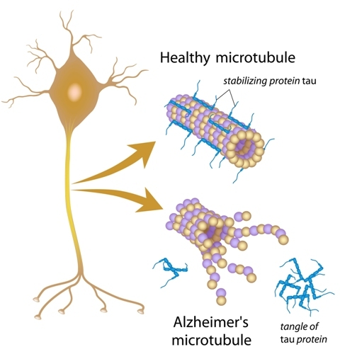
P-tau181 is a highly specific biomarker of AD and is a sub-type of misfolded tau protein. The occurrence of misfolded proteins can be triggered by genetics, environmental factors, or even head trauma to name a few. When a protein is misfolded it changes shape, leading to a functional change. Misfolded tau protein can also negatively change the shape of other correctly folded tau proteins, like a prion in Mad Cow disease, triggering neurofibrillary tangles (NFTs) to continue to aggregate and propagate down nerve networks interfering with neuronal functioning and causing cognitive decline in AD. 

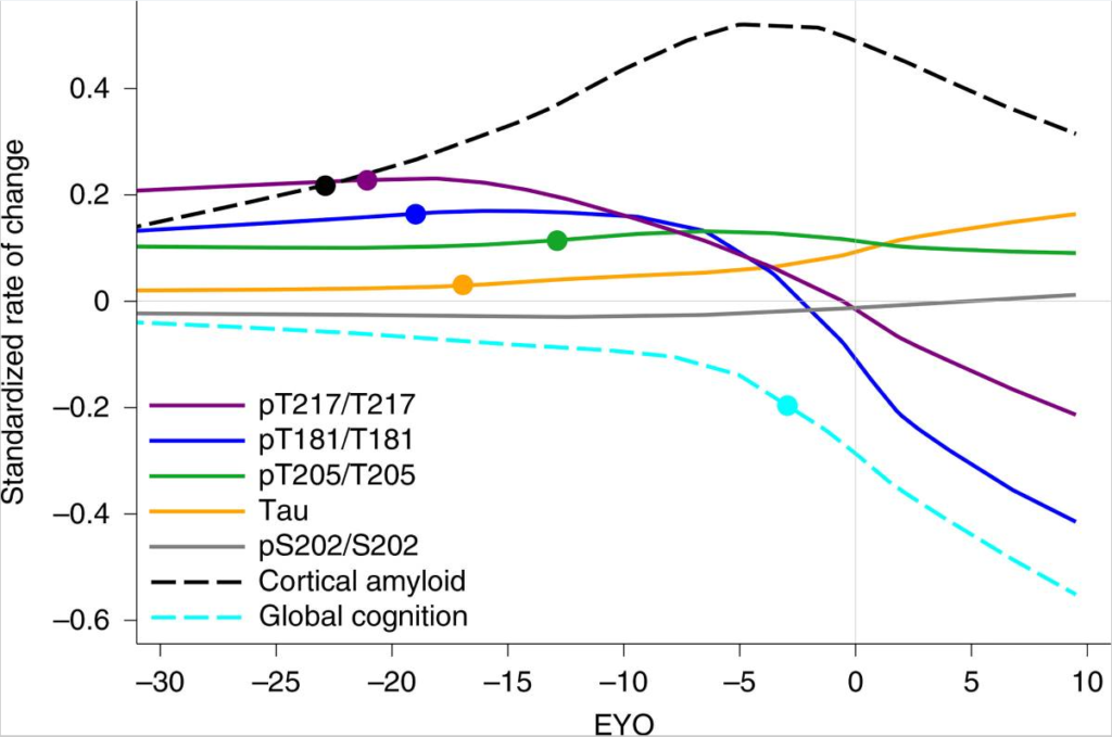
       Blood levels of p-tau 181 can differentiate AD from other neurodegenerative diseases, as well as predicting disease staging and the rate of cognitive decline. Subjects with AD were compared with cognitively unimpaired age-matched controls, patients with mild cognitive impairment (MCI), those with frontotemporal dementia and other neurodegenerative disorders, as well as healthy young adults. Established cerebrospinal fluid (CSF) and PET biomarkers were collected to compare the capability of blood p-tau181 for identifying AD.

Concentrations of serum p-tau181 significantly increased with cognitive decline across groups. The lowest p-tau181 concentrations were found in healthy young adults and cognitively unimpaired older adults. The next highest levels were found in amyloid β-positive cognitively unimpaired older adults and those with MCI. The highest concentrations were found in amyloid β-positive AD patients. Serum p-tau181 was not only sensitive in correctly identifying AD stages but also specifically ruled out other causes of dementia. 

A simple blood test would be invaluable for identifying and assessing AD in the community and clinical trials, especially since such p-tau181 concentrations correlate to AD risk. Here at the Center for Cognitive Health, we offer an AD prevention trial utilizing the p-tau 217 blood test, developed by Lilly, to assess the risk for developing AD in those with no memory problems–TRAILBLAZER-ALZ3 is using Donanemab (an antibody that targets amyloid-β), hopefully to prevent AD from developing. The days of needing a dose of radioactivity for an Amyloid PET scan or a spinal tap for CSF assessment may soon become obsolete. Hopefully, the results of this study will determine if treatment prevention (e.g., Donanemab) based on p-tau blood levels will be successful. If interested in knowing more about the study mentioned above, please visit our clinical trials page or give us a call at 503-207-2066 to find out more about disease modifying opportunities. 

Facebook
Twitter
Email
Sources
A donanemab prevention study in participants with Alzheimer’s disease (TRAILBLAZER-ALZ 3). (2022, April 7). ClinicalTrials.gov. Retrieved April 11, 2022, from https://clinicaltrials.gov/ct2/show/NCT05026866
Karikari, T. K., Pascoal, T. A., Ashton, N. J., Janelidze, S., Benedet, A. L., Rodriguez, J.L., Chamoun, M., Savard, M., Kang, M. S., Therriault, J., Schöll, M., Massarweh, G., Soucy, J. P., Höglund, K., Brinkmalm, G., Mattsson, N., Palmqvist, S., Gauthier, S., Stomrud, E., Zetterberg, H., … Blennow, K. (2020). Blood phosphorylated tau 181 as a biomarker for Alzheimer’s disease: a diagnostic performance and prediction modelling study using data from four prospective cohorts. The Lancet. Neurology19(5), 422–433. https://doi.org/10.1016/S1474-4422(20)30071-5

Exercise Related Neuroinflammatory Factor: Isolated

Previously, we’ve discussed ways in which we can lower our risk of Alzheimer’s Disease, such as decreasing risk for cardiovascular disease and eating a healthy Mediterranean-like diet. In this blog, we will dive deeper into the benefits of exercise and a particular protein upregulated from exercise, as there appears to be some implications in neurologic benefit regarding two major neurodegenerative disorders, specifically Parkinson’s Disease (PD) and Alzheimer’s disease (AD).

            Parkinson’s Disease is a disorder of the central nervous system that is characterized by impaired motor abilities including tremor, slowed and rigid mobility, unintentional movements, and even cognitive dysfunction (e.g., fluctuations in alertness, mild memory impairment). Regular physical activity is beneficial in slowing the progression of PD. In PD, neurodegeneration of the dopaminergic neurons in the substantia nigra occurs, damaging motor and reward systems.  Researchers compared PD patients who exercised to those who did not, and found that regular exercise slowed patient’s physical and cognitive decline but intermittent activity did not. Furthermore, exercise needed to be task and context specific to target declining functions in PD. For example, activities involved with balance-control, like Tai-Chi, better maintained postural and gait function compared to other activities. PD patients involved in household and work-related activities showed slower decline in activities of daily living (ADLs) and cognition. Additionally, regular physical activity increased corticostriatal plasticity and increased dopamine (DA) release stimulating increased activation of the striatum, possibly contributing to improved PD symptoms in those that habitually exercise. An encouraging takeaway from these findings suggests that something as simple as regularly sustained physical activity may lead to a modification in the typical trajectory of PD, slowing the rate of both the physical and cognitive decline, something that PD medications currently cannot accomplish. Furthermore, low levels of the brain protein clusterin predicts faster cognitive decline and dementia progression; clusterin increases with sustained exercise and may contribute to neurologic benefit.

A recent study compared the effects of physical activity with mice that regularly exercised to sedentary mice. After 28 days of running, the active mice, compared to their sedentary counterparts, showed increased overall cell survival in the memory circuit (called the hippocampus) as well as neural stem cells, progenitor cells, and astrocytes–all of which play important roles in neuronal maintenance, function, and repair. Similar beneficial results in the sedentary mice occurred with injection of serum taken from the exercised mice. When clusterin was removed from the exercised mice serum before injecting into the sedentary mice, the neurological benefits of decreased inflammation went away, supporting the role that clusterin may have in AD or PD when in lower proportions. To further understand these results, the researchers utilized RNA sequencing to analyze changes in gene expression in the active mice compared to their sedentary counterparts.  Physical activity in mice altered their gene expression, upregulating genes associated with hippocampal learning and memory. 

Humans with mild cognitive impairment (MCI) that exercise regularly for six months also show improved verbal and episodic memory, as well as significantly increased levels of clusterin. These results again indicate a role clusterin may play in AD and PD. 

All in all, exercise, and perhaps what is upregulated from exercise organically, like clusterin, may have a more powerful underlying effect on cognition than we knew previously. Such activity slowed decline in physical and cognitive functioning in PD, and may also work for those suffering from AD. Due to the increase in clusterin upon exercise, and the evidence presented above of clusterin’s capacity to improve brain function, one would wonder if providing clusterin may slow or prevent cognitive decline in those at risk. At this time, more research is needed to understand the potential implications of clusterin’s effect on cognition, especially in relation to diseases that affect cognitive functioning. Perhaps one day, for those of us who are too lazy to exercise, a clusterin pill will allow us to stay on the couch binge watching Netflix indefinitely! 

Facebook
Twitter
Email

Resources:

Hurley, D. (2022, February 17). A molecule transfers neurologic benefits of exercise to the sedentary. Neurology Today.

Mak, M.K.Y. & Schwarz, H.B. (2022). Could exercise be the answer? Neurology, 98(8), 303-304. doi:10.1212/WNL000000000001328

Tau: How Different Isoforms Predict Different Stages of AD Progression

       If you have read our blogs before, you are likely familiar with the two primary biomarkers of Alzheimer’s disease (AD), protein tau which forms neurofibrillary tangles (NFTs) and amyloid beta (Aꞵ) which forms amyloid plaques. Both of these contribute heavily to neuronal dysfunction, degeneration, and eventual memory impairment, but the relationship between them is complicated and has been the subject of research for several years. Evidence suggests that Aꞵ buildup instigates the misfolding of protein tau, eventually inducing NFT formation, however, tau levels better predict cognitive impairment than Aꞵ levels. More recently, researchers have expanded upon this by determining different stages of AD development as predicted by Aꞵ and tau.

       Before explaining the stages, we need to have some prior knowledge. While we frequently refer to tau as a single protein, this is not necessarily the case. Tau’s full name is microtubule associated protein tau (MAPT), and in its normal form it serves as the rigid scaffolding that helps maintain the shape of axons, the communication bridge between neurons. The diagram below depicts both normal, healthy tau as well as the NFTs that form in cases of AD. It is believed that the presence of toxic Aꞵ proteins induce hyperphosphorylation of tau proteins, changing their structure. This decreases their ability to support microtubules and makes them prone to clumping together, inducing dysfunction both through the tangle of proteins blocking normal cellular functions in the brain and through axonal loss due to their lack of stabilization.

       The specific locations on the protein at which tau can be hyperphosphorylated result in multiple different forms of tau, called p-tau isoforms. The most relevant isoforms to AD are p-tau217, 181, and 205. The presence, or lack thereof, of each type of tau predicts something different and generally correlates to a specific stage of disease progression. For example, an increase of p-tau217 and 181 without presence of NFTs predicts amyloidosis, the buildup of Aꞵ plaques in the brain before symptom onset. A rise of p-tau205 as measured by cerebrospinal fluid (CSF) correlates to waning brain metabolism and shrinking gray matter, the initial stages of degeneration but not yet producing dysfunction. Finally, as total tau levels spike in CSF, NFTs begin to form and cognitive decline begins. Interestingly, once NFT formation begins and global cognition starts to decline, the amount p-tau181 and 217 present in CSF plummets, presumably because these isoforms are being sequestered into the NFTs that are now forming. While this explanation for decrease in CSF p-tau levels is hypothetical, it is supported by the fact that the same phenomenon occurs with amyloid. The figure below from Barthélemy et. al. (2020) exemplifies this sudden change in p-tau and amyloid levels around the estimated year of onset (EYO).

       This information is extremely useful because AD therapies being tested in clinical trials utilize many different mechanisms to fight the disease. Using the different p-tau metrics above, it may be possible to more specifically gauge how far progressed a patient may be and what therapies are most likely to be useful. It is also projected that the increased specificity for placement in trials provided by p-tau measurements, as well as tau PET scans using a new and more accurate tracer, could reduce the sample size needed within clinical trials to find (or disprove) efficacy. Specifically, for trials on preclinical (asymptomatic) AD, using p-tau217 with tau PET scans was hypothesized to reduce required sample size by 43% and by 68% for MCI trials. Using either p-tau217 or tau PET alone would theoretically also result in reduced sample requirements, albeit to a lesser degree, with p-tau217 alone for preclinical AD trials reducing sizes by 31%, and PET alone reducing MCI trial sizes by 47%.

       A decreased sample size with more specific subject selection could provide faster clinical trial outcomes with lessened screening times, and a decrease in the likelihood of a successful drug requiring additional data before coming to market. Should these staging procedures become a widespread method of pre-screening, patients are more likely to be placed into a clinical trial that will help them based upon their specific disease staging, whether that be clearing tau tangles, preventing tau aggregation, or clearing amyloid proteins before they even initiate the hyperphosphorylation of tau.

Facebook
Twitter
Email
Sources:
Different CSF Phospho-Taus Match Distinct Changes in Brain Pathology. Alzforum [Internet]. 2020. Available from: https://www.alzforum.org/news/research-news/different-csf-phospho-taus-match-distinct-changes-brain-pathology
In Preclinical Alzheimer’s, p-tau217 in Blood Best Predicts Tangles. Alzforum [Internet]. 2021. Available from: https://www.alzforum.org/news/research-news/preclinical-alzheimers-p-tau217-blood-best-predicts-tangles
Barthélemy et. al. A soluble phosphorylated tau signature links tau, amyloid and the evolution of stages of dominantly inherited Alzheimer’s disease. Nature Medicine [Internet]. 2020. Available from: https://www.ncbi.nlm.nih.gov/pmc/articles/PMC7309367/

Insulin Resistance and Alzheimer’s: A Two Way Street (and How GLP-1 Receptor Agonists May Help Cross It)

       Previously, we described the relationship between insulin resistance and AD, and treatments pertaining to such (https://www.centerforcognitivehealth.com/insulin-and-ad/). However, the overarching principle of how insulin signaling ties into development of neurodegenerative conditions is only loosely understood, indicating the need for further research.

       Insulin, produced by the pancreas, signals to maintain glucose homeostasis and cell growth/survival by binding to insulin receptors (IRs). Insulin resistance is caused by a downregulation of these IRs, which in turn instigates an overproduction of insulin (hyperinsulinemia) to try to overcome the limited signaling. IRs are present in large quantities in the brain, especially in the hippocampus, a prominent structure for memory. During hyperinsulinemia episodes our bodies downregulate the transporters that allow insulin into the brain, possibly increasing cell death, decreasing cell growth, and impairing memory. Diseases, such as AD and Parkinson’s disease (PD), are twice as likely to develop in individuals with diabetes, supporting this relationship.

       While diabetes increases the risk of AD, AD also increases the risk of developing type II diabetes mellitus (T2DM). Research into AD’s role in causing T2DM showed that toxic amyloid-ꞵ (Aꞵ) oligomers in the AD brain interact with hippocampal tissues to reduce the number of IRs present, and is predictive of insulin resistance outside the brain, eventually inducing T2DM. Furthermore, inflammation is strongly tied to the development of both T2DM and AD, possibly explained by the fact that insulin resistance increases circulating inflammatory cytokines.

       It appears that treating peripheral insulin resistance has both a direct and indirect impact on risk/prevention of AD on top of the obvious impact on diabetes/insulin resistance. Clinical trials aimed at treating AD have taken notice. For instance, we currently have a trial utilizing semaglutide, a medication already approved as an antidiabetic treatment, to attempt to stop/slow progression of AD in individuals with Mild Cognitive Impairment or Early AD. A hormone called GLP-1 has also been implicated in playing a role in both diabetes and AD. GLP-1 is similar to insulin with a strong role in glucose homeostasis but is quickly degraded under normal circumstances. Semaglutide, a GLP-1 receptor agonist (RA), simulates the effects of GLP-1 while avoiding quick degradation, creating lasting impacts on glucose regulation without being impacted by insulin resistance.

       Before semaglutide, several other molecules were tested for this purpose. The first GLP-1 RA, exendin-4, improved cognition and reduced Aꞵ presence in the brains of both AD mice and wild-type mice. The next major GLP-1 RA, liraglutide, produced longer lasting effects than exendin-4 and was shown to prevent Aꞵ neurotoxicity and reduce Aꞵ plaques in the hippocampus and cortex, reduce cell death, alleviate brain insulin resistance, and improve memory in the same mouse model exendin-4 was tested on. It also lowered levels of phosphorylated Tau, the other major protein implicated in AD progression. When administered before significant plaque burden was present and memory impairment began, liraglutide slowed disease progression in AD mouse models. Yet another marketed diabetes drug, lixisenatide, enhances long-term potentiation, and lowers Aꞵ plaque load, microglial activation, and neurofibrillary tangles. Despite these other treatments, semaglutide shows the greatest effectiveness compared to other GLP-1 RAs with regards to glycemic regulation. Given how intertwined insulin resistance and neurodegeneration seem to be, greater efficacy in one instance may offer benefit in the other. Furthermore, semaglutide is already approved and marketed for treatment of diabetes so it’s safety and tolerability are well studied.

       With all this therapeutic potential surrounding semaglutide and GLP-1 RAs, if you or someone you know between the ages of 55 and 85 is experiencing Mild Cognitive Impairment (MCI) or mild Alzheimer’s dementia they may be eligible to screen for the trial! Feel free to contact our office or inquire about potential involvement on our website.

Facebook
Twitter
Email
Sources:
Batista, A.F., Bodart-Santos, V., De Felice, F. G., & Ferreira, S.T. Neuroprotective Actions of Glucagon-Like Peptide-1 (GLP-1) Analogues in Alzheimer’s and Parkinson’s Diseases [Internet]. CNS Drugs. 2018. Available from: https://www.researchgate.net/publication/329373799_Neuroprotective_Actions_of_Glucagon-Like_Peptide-1_GLP-1_Analogues_in_Alzheimer’s_and_Parkinson’s_Diseases
Insulin and Alzheimer’s Disease [Blog]. 2020. Available from: https://www.centerforcognitivehealth.com/insulin-and-ad/
Alsugair, H.A., Alshugair, I.F., Alharbi, T.J., Bin Rsheed, A.M., Tourkmani, A.M., Al-Madani, W. Weekly Semaglutide vs. Liraglutide Efficacy Profile: A Network Meta-Analysis [Internet]. Healthcare. 2021. Available from: https://pubmed.ncbi.nlm.nih.gov/34574899/

TREM2, Microglia, and Alzheimer’s: The Final Puzzle Piece?

       Alzheimer’s researchers have spent years focusing on the “amyloid hypothesis” proposing that toxic ꞵ-amyloid (Aꞵ) proteins, and the plaques they form outside neurons, were the primary cause of Alzheimer’s disease (AD). However, even therapies that successfully cleared Aꞵ in symptomatic individuals failed to slow or stop neurodegeneration, suggesting something else may be at play. Further research showed that Aꞵ buildup instigates the dysregulation of tau protein forming neurofibrillary tangles (NFTs) that aggregate inside neurons. When NFTs form near amyloid plaques the tau proteins may become detached, causing neuronal dysfunction. In this form, they are called neuritic plaques. On top of these complex processes, an intermediate step between Aꞵ build-up and the formation of neuritic plaques mediated by microglia may exist.

       Under normal circumstances, microglia are the brain’s immune cells and support neurons to combat pathogens and repair injured tissues by absorbing/breaking down debris, excess synapses, and pathogens. In the case of AD, one study found that microglia surround Aꞵ plaques and interact with the fibers, potentially preventing further aggregation or limiting the effects of toxic Aꞵ, supported by another study that found that fewer microglia around plaques predicted greater damage to nearby axons. Several AD risk-factor genes are expressed predominantly in microglia as opposed to neurons. For example, the R47H variant of the TREM2 gene, which codes for a microglial receptor, seems to impair microglial function and predicts more neuritic plaques and axonal damage. When the TREM2 gene was completely knocked out in mice with amyloid pathology microglia suffered major functional deficits, inhibiting them from interacting with amyloid plaques and leading to an increase in swollen, damaged neurons.

       If this R47H variant impairs microglial function thereby increasing neurodegeneration, then improving microglial function should be protective. Unfortunately, the answer is not so cut and dry. In mice with amyloid pathology but no tau pathology, this theory holds true and microglial activity seems to protect the brain. However, once tau pathology presents in mice, microglia modify their genetic expression activating genes that synergize with the presence of the APOE4 gene and are associated with greater neurodegeneration. To confirm this, researchers knocked out the TREM2 gene in mice with tau pathology and, unlike the previous knockout experiment with only amyloid pathology, this TREM2 knock out decreased neurodegeneration. In another study, mice with early tau pathology had their APOE4 levels reduced by half, resulting in decreased microglial activation and reduced neuronal damage.

       As these opposing results based on disease-phase show, the role of microglia in AD neurodegeneration (at least in mice) seems to change depending upon the presence or lack of tau pathology. To understand why this is, we must remember that microglia are immune cells that use cytokine signaling to communicate with other immune cells and mediate inflammatory responses. Research suggests that cytokines released by microglia exacerbates tau dysfunction and begin NFT aggregation. Evidence indicates that microglia have an important role in fighting amyloid buildup and pathology, protecting the brain in the early disease stages, but become detrimental once tau pathology begins as they overreact to the presence of neuritic (tau positive) plaques, increasing neurodegeneration and inflammation in response to the presence of misfolded tau.

       As such, researchers have begun to factor microglial activation per disease-stage into potential treatment models. Some suggest that it may be beneficial to up-regulate activation of microglia in the initial stages of amyloid build-up, allowing them to surround the plaques and prevent propagation of the pathology as long as possible. In fact, one such therapy is already being developed and tested for efficacy in clinical trials! AL002, developed by Alector, Inc., is a TREM2 agonist expected to upregulate microglial activity around amyloid plaques in the early disease stages prior to significant tau pathology, thereby slowing the rate of symptom onset and neurodegeneration. Excitingly, this trial will be using our facility as a research site so if you or someone you love is experiencing mild cognitive impairment or early Alzheimer’s disease, contact our office and we can discuss your eligibility for the trial. However, once tau pathology starts to develop, treatments would theoretically have to target inactivation of microglia in order to prevent their destructive effects on neurons via cytokine interaction with neuritic tau plaques, but no trials are yet testing this hypothesis.

Facebook
Twitter
Email
Sources:
       Ulrich, J. & Holtzman, D.M. A New Understanding of Alzheimer’s [Magazine]. Scientific American. 2021.
       Gratuze, M., Leyns, C.E., & Holtzman, D.M. New insights into the role of TREM2 in Alzheimer’s disease [Online]. Molecular Neurodegeneration. 2018. Available from: https://molecularneurodegeneration.biomedcentral.com/articles/10.1186/s13024-018-0298-9.
       Mrak, R.E., Sheng, J.G., & Griffin, W.S. Glial cytokines in Alzheimer’s disease: review and pathogenic implications [Online]. Human Pathology. 1995. Available from: https://pubmed.ncbi.nlm.nih.gov/7635444/.

Donanemab Research Links Amyloid and p-tau217

       The FDA approval of Biogen’s aducanumab, now known as Aduhelm, set a low bar that other potential antibody treatments for Alzheimer’s disease are also hopeful to meet. Researchers from Eli Lilly recently reported that donanemab administered at plaque-dissolving strength correlated with diminishing levels of plasma p-tau217. When their data was plugged into a disease-progression model, it suggested that the decrease in amyloid and tau correlated with slower cognitive decline. Similarly, the Phase 2 trial and open-label extension of Biogen/Eisai’s lecanamab provided evidence of the drug’s disease modifying effect. The FDA has granted both donanemab and lecanamab breakthrough therapy status, and both Lilly and Biogen/Eisai have announced they will be using their Phase 2 data to seek accelerated approval.

       The most recent analysis of donanemab was carried out using data from Lilly’s TRAILBLAZER trial. During this trial, 131 participants randomized into the treatment group received infusions each month, with a stable dose of 1,400 mg after the first three half-doses at 700 mg. Once the participant’s amyloid burden fell below 25 centiloids, the dose was lowered to 700 mg. If the amyloid burden remained below 25 for two consecutive scans or if it fell below 11, participants were switched to placebo. By the end of the 76-week trial, results showed a 32 percent slowing of decline on the Integrated Alzheimer’s Disease Rating Scale (iADRS). Participants who cleared amyloid below 11 centiloids and were switched to placebo by 24 weeks showed amyloid burdens barely above 11 centiloids at 76 weeks. For this group, it would take 14 years for amyloid to accumulate back to baseline levels of approximately 90 centiloids. They also show reduced tangle burden in the temporal, parietal, and frontal lobes compared to participants in the placebo group.

       Several sources have reported that p-tau217 presence in CSF and plasma correlates with disease trajectory and amyloid and tangle burden. After 12 weeks of donanemab treatment, plasma p-tau217 fell significantly from subject-matched baseline measurements. This trend continued and strengthened in statistical significance during each follow-up assessment. At the end of the trial, the placebo group had a 6 percent increase in plasma p-tau217, while this level had decreased 24 percent from baseline in the treatment group. The drop in p-tau217 strongly correlated with a reduction in amyloid and tau tangles, suggesting that neurons release phospho-tau when amyloid is present. Thus, plasma p-tau217 may be used as biomarker to assess the effects of novel treatments on beta-amyloid fibrils. If its clinical efficacy is confirmed, it will be greatly useful in the development of Alzheimer’s treatments.

       Overall, the most intriguing implication of the donanemab data is that patients may only need to be treated for a few months to see lasting results. The link between amyloid and p-tau217 means the results could be easily and inexpensively tracked due to the simplicity of access to plasma, compared to current ways of testing amyloid levels with PET scans or spinal taps. Furthermore, this would be the strongest evidence that amyloid deposition induces tangle formation. If donanemab receives FDA approval, it may soon become a common treatment for seniors experiencing memory problems and even those preclinical patients, who are cognitively normal but at risk for developing Alzheimer’s disease. As Eli Lilly, Biogen, and Eisai begin the process of fast-track approval, we will likely know more in the coming months.

Facebook
Twitter
Email
Source: 
On Donanemab, Plaques Plummet. Off Donanemab, They Stay Away. (2021, August 6). ALZFORUM. https://www.alzforum.org/news/conference-coverage/donanemab-plaques-plummet-donanemab-they-stay-away

The Case for the Amyloid Hypothesis

       The amyloid hypothesis has inspired most of Alzheimer’s research for the last 20 years. It proposes that accumulation of a protein fragment known as beta-amyloid, and in particular the 42 amino acid subtype, is the underlying cause of the Alzheimer’s pathogenesis. It is a “sticky” compound that builds up within the brain 10-15 years before symptoms emerge. In theory, it disrupts synaptic communication, and ultimately ends in neuronal death by initiating neural fibrillary tangle (NFT) formation. The theory posits overproduction, poor disposal, and eventual accumulation of beta-amyloid is the primary cause of Alzheimer’s disease. 

       Beta-amyloid is only a small section of the larger amyloid precursor protein (APP). At present, scientists have not determined the normal function of APP. However, they know that when the protein is activated, it is divided into smaller pieces that are both inside or outside of cells. When pieces of beta-amyloid begin to accumulate, they form small clusters called oligomers. Multiple recent studies have implicated beta-amyloid oligomers as upstream drivers of Alzheimer’s disease. As these oligomers accumulate in the brain, they create chains of clusters known as fibrils, which then bond together to form beta-sheets. The characteristic amyloid plaques found in Alzheimer’s patients are composed of beta-sheets meshed together with other substances, such as tau protein making up NFTs. These plaques are believed to create intra-neuronal NFTs that cause synaptic injury, loss of neuronal function, and eventually cell death.

       Genetic studies of familial Alzheimer’s patients identified over 200 mutations that lead to an early onset of the disease. Many of these mutations occur in the APP gene and cause an increase in the production of beta-amyloid. Overproduction causes an increase in misfolded beta-amyloid, resulting in the formation of amyloid oligomers and eventually plaques.

       The APOE4 genotype has been identified as the greatest risk factor for developing Alzheimer’s disease. Those who carry the E4 allele of apolipoprotein E (APOE) have a much greater risk for developing early onset Alzheimer’s disease. The APOE4 genotype, which has been identified in approximately 65% of Alzheimer’s patients, is associated with reduced ability to clear amyloid from the brain and increased aggregation of beta-amyloid into oligomers. 

     Another argument for the amyloid hypothesis comes from examining the brains of individuals with Down syndrome. Adults with Down syndrome have three copies of the chromosome carrying the APP gene: chromosome 21. These individuals invariably develop beta-amyloid plaques identical to those seen in Alzheimer’s disease and have a high risk of developing early onset dementia. Thus, overproduction of beta-amyloid plaques in Down syndrome results from a comparable genetic pathology to Alzheimer’s patients with mutations of the APP gene.

       Clinical trials of anti-amyloid agents that target beta-amyloid oligomers, such as aducanumab, donanemab, and lecanemab, have exhibited statistically significant clinical efficacy. Though as discussed in previous blog posts, the benefit received from these drugs is directly related to how early they’re given in the progression of the disease, generally before amyloid plaques have accumulated to the point of causing too much neural damage, which suggests that beta-amyloid accumulation initiates the cascade of Alzheimer’s pathogenesis. 

       The genetic and clinical trial data suggests beta-amyloid plays a key part in Alzheimer’s disease. The research sparked by the amyloid hypothesis remains relevant to this day and continues to inspire further clinical investigation. However, there is further emerging evidence that suggests it is far from the only factor influencing pathogenesis. Future studies targeting beta-amyloid and further examining APP mutations may one day lead to promising therapeutic interventions for Alzheimer’s disease, but alternative methods should also be pursued. Check in next week when we’ll be discussing these alternative methods.

Facebook
Twitter
Email
Sources:
Annus, T., Wilson, L. R., Hong, Y. T., Acosta–Cabronero, J., Fryer, T. D., Cardenas–Blanco, A., Smith, R., Boros, I., Coles, J. P., Aigbirhio, F. I., Menon, D. K., Zaman, S. H., Nestor, P. J., & Holland, A. J. (2016). The pattern of amyloid accumulation in the brains of adults with Down syndrome. Alzheimer’s & Dementia, 12(5), 538–545. https://doi.org/10.1016/j.jalz.2015.07.490
Beta-amyloid and the Amyloid Hypothesis. https://www.alz.org/national/documents/topicsheet_betaamyloid.pdf
Tolar, M., Hey, J., Power, A., & Abushakra, S. (2021). Neurotoxic Soluble Amyloid Oligomers Drive Alzheimer’s Pathogenesis and Represent a Clinically Validated Target for Slowing Disease Progression. International Journal of Molecular Sciences, 22, 6355. https://doi.org/10.3390/ijms22126355


Why has Biogen’s aducanumab become so controversial?

The FDA’s approval of aducanumab has been shrouded by controversy, with experts quickly criticizing the FDA’s ruling, referencing two large studies showing little convincing evidence of efficacy. These feelings were mirrored by the FDA’s own advisory committee. Of the 11 members, 10 voted against the drug’s approval, citing insufficient evidence, and one member was undecided. The drug was approved anyway, and three committee members have since resigned.

To understand the reasoning behind their actions, let’s discuss the science behind the drug’s approval. Biogen’s aducanumab, now marketed under the name Aduhelm, is an amyloid antibody that works to clear β-amyloid from the brain. Previous drugs have successfully cleared amyloid from the brain but did not improve cognitive function. Most of Biogen’s trials produced similar results.

Only after re-analysis was Biogen able to show cognitive decline slowed in a subset of patients from their EMERGE trial. The positive effects were seen only in patients who carried the APOE4 genotype, while patients who were not carriers fared worse on the drug than the placebo. The identical ENGAGE trial failed to produce positive results altogether.

The negative results from the ENGAGE trial are important because the positive effects seen in APOE4 patients from the EMERGE trial only began to occur following a change in study protocol. Initially, APOE4 patients were receiving lower doses of the drug because they are more prone to amyloid-related imaging abnormalities (ARIA) while receiving higher doses. Midway through the trial, this was adjusted so that all patients were receiving the same dose per kilogram. This caused more APOE4 patients to experience imaging abnormalities, making it necessary to pause dosing and MRI monitoring until the condition cleared. Therefore, many of these patients and their physicians became aware that they were likely receiving the drug, breaking the blind and possibly resulting in bias data.

Biogen maintains that the increased dosage produced greater efficacy in this subset of patients following the change in protocol and used this data to obtain FDA approval. However, positive results may have stemmed from the unintentional unblinding of the treatment group, potentially affecting ratings on measures of cognitive function and daily living. Thus, the drug was approved based on potentially erroneous data. 

An independent analysis of the heterogeneity of Alzheimer’s disease progression points to further issues with the EMERGE and ENGAGE results. When comparing individual trajectories on cognitive assessments, performance was highly variable in prodromal and mild Alzheimer’s disease patients. When these results were compared to the data from actual clinical trials (i.e. Biogen’s EMERGE and ENGAGE), the differences fell within the range expected when there is no treatment effect. Therefore, it’s also important to consider that the results generated by Biogen could be due to oversampling of individuals who are declining faster in the placebo group or declining slower in the treatment group.

Following aducanumab’s approval, it is unclear if it will become the new standard that potential new treatments are compared to instead of placebo. What we do know now is that Biogen will be required to conduct a new randomized controlled phase 4 trial to verify the efficacy of aducanumab. During the first two years, Biogen will have a monopoly on the market and participants will be required to pay approximately $56,000 upfront as it stands now, although some supplemental insurances may reduce this rate. Even if Medicare does fully reimburse, the burden on the system would be significantly higher than any other drug currently on the market. If they decide not to cover the cost, very few people will be able to utilize it.

Moreover, the drug relies heavily on the hypothesis that amyloid is the underlying cause of Alzheimer’s disease. If a patient has received an amyloid PET scan and knows their APOE status, then the drug may be worth it in the early stages of the disease. However, if the disease has progressed too far, removing amyloid doesn’t translate to improved cognition or functioning.

All in all, whether aducanumab is worth it depends on the patient and the situation. If you’re in the early stages of the disease, know your APOE status, have had an amyloid PET scan, and are willing to pay upfront for treatment, it may be worth your while. However, few people fall into this category. Our hope is that research will continue to explore other options independently and without comparison to Biogen’s aducanumab.

Facebook
Twitter
Email
Sources:
https://n.neurology.org/content/96/22/e2673
https://time.com/6072980/alzheimers-drug-approval-controversy/
https://www.alzforum.org/news/research-news/aducanumab-still-needs-prove-itself-researchers-say#enlarge
https://www.kff.org/medicare/issue-brief/fdas-approval-of-biogens-new-alzheimers-drug-has-huge-cost-implications-for-medicare-and-beneficiarie

Donanemab: A Promising Anti-Amyloid Therapy

     Due to inconsistent effects of anti-amyloid therapies, such as aducanamab, a recent shift from the “amyloid hypothesis” to the “tau hypothesis”, has occurred. The primary issue with most anti-amyloid therapies, is that the successful removal of amyloid-β (Aβ) plaques doesn’t significantly reduce the clinical symptoms, suggesting that significant damage to the underlying brain structures has already occurred. However, donanemab, another anti-amyloid therapy, promises to be different, and here’s how.

        Firstly, donanemab, unlike most of the previous anti-Aβ therapies, is designed for early use in Alzheimer’s disease (AD), when Aβ levels are notable yet phosphorylated tau (pTau) making up neurofibrillary tangles (NFTs) are not widely deposited. The presence of amyloid is believed to form pTau misfolding and aggregation into NFTs. Not including individuals with widely dispersed NFTs helped provide a clearer picture of what Aβ removal can improve, without significant NFT presence contributing to continued dysfunction and cell death. Secondly, in a Phase-II trial donanemab met its primary outcome measure, producing a significant improvement in 2 cognitive/functional rating scales compared to controls. The secondary outcomes did not reach significance in this trial, but likely could have in a trial with more participants (such as the stage 3 trial with donanemab that our site is participating in). Finally, it targets a specific type of Aβ known as N3 truncated, pyroglutamylated Aβ (pE-Aβ) which is present in relatively low quantities compared to other types of Aβ but is especially prone to “seeding” or propagation of Aβ pathology by interacting with and misfolding other proteins.

      This pE-Aβ has been researched previously and is strongly implicated in the pathogenesis of AD. Its role in neurodegeneration was confirmed in AD mouse when its inhibition improved memory and decreased Aβ deposition. This is because pE-Aβ works synergistically with conventional Aβ when co-incubated, forming resilient hybrid oligomers that have stronger cytotoxic effects than oligomers formed by non-pE-Aβ alone. Therefore, a drug such as donanemab that targets pE-Aβ before they initiate the cascade of plaque formation could, in theory, prevent significant plaque-induced neurodegeneration.

         Even more exciting, in the Phase-II trial also improved Aβ clearance with spectacular results such that within 24 weeks of starting treatment, 25% of the subjects on active drug reached Aβ negativity as reflected in Amyloid-PET scans. By week 76, 68% were Aβ negative. Furthermore, as expected, these results were strongest in those with lower pTau burden at baseline. On the other hand, there were also unexpected outcomes such as the lack of significant hippocampal volume change and an overall decrease in brain volume and increase in ventricular size for those in the treatment group compared to controls. Researchers hypothesized this was due to the rapid removal of plaque volume. Despite these promising outcomes, donanemab is not expected to be a sole treatment for AD in the future. Alzheimer’s is a multi-factorial disease and therefore researchers expect to someday utilize a multi-factorial combination of therapies to specifically target each phase of the disease spectrum and the new Phase-III TRAILBLAZER-ALZ2 trial for donanemab may emerge as one of these future treatments!

Facebook
Twitter
Email
Sources:
Talan, J. A New Monoclonal Antibody Shows Promise Early in Alzheimer’s Disease [Article]. Neurology. 2021.
Nussbaum, J., et al. Prion-like behavior and tau-dependent cytotoxicity of pyroglutamylated amyloid-β [Online]. Nature. 2012. Available from:  https://www.ncbi.nlm.nih.gov/pmc/articles/PMC3367389/

Lion’s Mane: A Mushroom to Remember

       This week we will be discussing the mushroom Lions Mane, or Hericium Erinaceus, and its health applications for Alzheimer’s Disease (AD). Currently, there are no drugs on the market that prevent, reverse, or halt AD progression. Although a few clinical trials in the pipeline show promise, scientists are also looking to alternative treatments, like Lions Mane, to combat AD.

       Lions Mane is a culinary mushroom and is commonly eaten in countries such as Japan and China without any harmful effects. It’s generally found growing under old broadleaf trees and contains erinacines, natural substances with potential pharmacological effects on the central nervous system (CNS). There are two forms of Lions Mane that can be ingested; the fruiting body and the mycelium that encompasses the erinacines. Erinacines fall within a group of compounds called cyathin diterpenoids, and are stimulators of nerve growth factors (NGF). Nerve growth factors play a supportive role in the CNS, and are critical to adequately protect surviving and developing neurons.

       Rats given erinacine A for 3 weeks increased their concentrations of noradrenaline and homovanillic acid in the hippocampus compared to controls and showed evidence of an overall increase in NGF levels, most notably in the dentate gyrus of the hippocampus. Increases in noradrenaline and homovanillic acid result in more alertness and better retrieval of memory along with breaking down fat and increasing blood sugar levels to promote more energy, suggesting that erinaceus A promotes nerve and brain health in animal models. Furthermore, H. erinaceus mycelium containing erinacine A administered orally to AD transgenic mice for 30 days resulted in decreased recruitment and activation of plaque burden compared to controls. Other structures often impaired in AD, like the hippocampus and the locus coerulous, also showed functional improvement compared to those not given the mycelium.

       A double-blind clinical trial assessing the oral administration of H. erinaceus fruiting bodies in elderly humans showed improvement in subjects with mild cognitive impairment compared to age-matched controls. Researchers measured improvements using the Revised Hasegawa Dementia Scale (HDS-R). The group ingesting H. erinaceus significantly increased their scores during the 16-week treatment period, indicating improvement compared to those not taking H. erinaceus. However, when subjects stopped taking H. erinaceus their scores began to fall, reflecting scores similar to those that were untreated, indicating the need for continued use.

       Several different compounds in H. erinaceus appear to contain protective benefits, such as amyloid plaque reduction, insulin-degrading enzyme expression, enhancing NGF release, and even managing neuropathic pain. Although some is known about erinacines, many of the compounds are still undergoing research, with some still being discovered. These discoveries and continued ones will hopefully continue to pave the way for therapeutic strategies to prevent, manage, and slow AD progression. Furthermore, prior research and clinical trials have proven that Lion’s Mane and its extracts are safe for human consumption at doses of 3-4 grams per day (although allergies have been noted). If this research interests you, we encourage you to discuss Lion’s Mane with your primary care physician to potentially improve your cognition.

Facebook
Twitter
Email
Sources:
Li, I. C., et al. NeuroHealth Properties of Hericium erinaceus Mycelia Enriched with Erinacines [Online]. 2018. Available from: https://pubmed.ncbi.nlm.nih.gov/29951133/
Mori, K., et al. Nerve Growth Factor-Inducing Activity of Hericium erinaceus in 1321N1 Human Astrocytoma Cells [Online]. 2008. Available from: https://pubmed.ncbi.nlm.nih.gov/18758067/
Chong, P. S., Fung, M. L., Wong, K. H., & Lim, L. W. Therapeutic Potential of Hericium erinaceus for Depressive Disorder [Online]. 2019. Available from: https://pubmed.ncbi.nlm.nih.gov/31881712/
Mori, K., Inatomi, S., Ouchi, Y., Azumi, Y., Tuchida, T. Improving effects of the mushroom Yambushitake (Hericium erinaceus) on mild cognitive impairment: a double-blind placebo-controlled clinical trial [Online]. 2009. Available from: https://pubmed.ncbi.nlm.nih.gov/18844328/
Shimbo, M., Kawagishi, H., Yokogoshi, H. Erinacine A increases catecholamine and nerve growth factor content in the central nervous system of rats [Online]. 2005. Available from: https://www.sciencedirect.com/science/article/abs/pii/S0271531705001041
Chen, C., et al. Erinacine S, a rare sestererpene from the mycelia of Hericium erinaceus [Online]. 2016. Available from: https://pubmed.ncbi.nlm.nih.gov/26807743/