The French Paradox: Red Wine for a Healthy Mind?

Known since 1992, the French diet is high in saturated fats, a risk for coronary heart disease (CHD), yet they have less than half the CHD-related deaths compared to the US, Sweden, and the UK. High intake of wine, thought to be 57% of alcohol consumption in France, may contribute to this disproportionately low frequency of CHD representing the French paradox. However, heavy alcohol consumption is associated with increased risk of heart attack, arrhythmia, hypertension, and sudden death. This raises the question; How does wine consumption improve cardiovascular health?

Wine, particularly red wine, contains high levels of phenols. One phenol, resveratrol, may contain cardiovascular protectant properties. It inhibits oxidative stress caused by free radicals, preventing cell damage or death. Resveratrol appears to increase lifespan and promote healthy aging. Fruit flies, fish, and nematodes given resveratrol increase their lifespan significantly! In humans low to moderate amounts of wine consumption are associated with decreased cardiovascular- and cerebrovascular disease-related deaths.

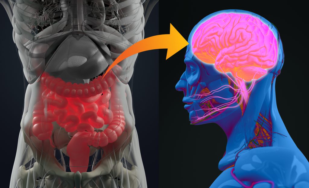
Moderate consumption of wine is also associated with lower instances of Alzheimer’s disease (AD). Normally, proteasomes are responsible for degradation of damaged and aggregated proteins such as Aβ, but their activity is impaired in AD. Oxidative stress inactivates proteasomes, which can be prevented with resveratrol administration in disease-model cell cultures. Administration of resveratrol in vitro correlates to increased intracellular degradation of Aβ by proteasomes, suggesting that moderate wine consumption may decrease one’s likelihood of developing AD. Synthetic resveratrol supplements are new to the field and require further research.

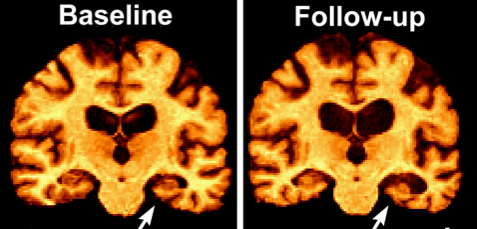
One year of resveratrol supplementation (500-2000 mg per day) slowed decline in cognition and function compared to placebo. Yet other studies found no difference with supplementation over 52 weeks. Larger studies over a longer duration are needed. Pterostilbene, a synthetic resveratrol analog, has much higher oral bioavailability and blood-brain barrier permeability warranting further research. Resveratrol administration correlates with decreased central nervous system (CNS) deposition of Aβ, and increased brain shrinkage in AD patients as a product of reduced neuroinflammation.

Resveratrol benefits a variety of other physiological functions, too. It delays or prevents cell death in a variety of cell types, decreases atherosclerotic lesion formation, reduces risk for hypercholesterolemia, maintains glucose homeostasis in diabetes, and promotes tumor suppressor gene expression. In rat models of Lewis lung carcinoma, resveratrol decreases tumor size, weight, and metastasis, indicating a diverse range of effects on chemoprevention. It has powerful effects on energy metabolism. In mice, administration increased aerobic capacity as evidenced by increased running time and oxygen consumption in muscle fibers. Its effects on energy metabolism might also minimize damage from secondary spinal cord injuries. Further research in human models is needed to validate it as a therapeutic.

While resveratrol’s impacts on cognition and AD are inconclusive, it has potential to benefit health in a variety of other ways, which may justify a glass of red wine every so often. If you can’t drink wine, resveratrol is also present in a number of foods, including grapes, peanuts, soybeans, apples, and pomegranates. Red wines contain concentrations between 0.361-1.972 mg/L, meaning that one would have to drink many bottles of wine to achieve the hypothesized therapeutic dose (TD) of 1 gram per day. Even including resveratrol containing foods such as peanuts (0.03-0.14 μg/g) and apples (400 μg/kg) does not reach the TD.  However, these measurements only account for unbound resveratrol. Food and drinks containing pure resveratrol also contain molecular constituents and resveratrol glucosides which occur in higher concentrations and, in some cell culture and animal studies, show higher potency than resveratrol itself. These molecules may actually be the driving force behind the French Paradox, but focused research and clinical trials will be required to confirm this hypothesis.

There are also supplemental tablets derived from Japanese knotweed containing a therapeutic dose of concentrated resveratrol. Unfortunately, research has shown that these supplements are a less effective source of resveratrol as it’s bioavailability and absorption is enhanced by the food matrix present in its naturally occurring forms. Regardless, with so many beneficial impacts in the body and no serious adverse effects we could all stand to increase our resveratrol intake – whether it comes from a glass of red wine, a handful of peanuts, or a supplement. This week, go out and live like the French!

Facebook
Twitter
Email
Sources: 
Renaud, S., & de Lorgeril, M. Wine, alcohol, platelets, and the French paradox for coronary heart disease [Internet]. Epidemiology. 1992. Available from: https://www.thelancet.com/journals/lancet/article/PII0140-6736(92)91277-F/fulltext
Catalgol, B., Batirel, S., Taga, Y., & Kartal Ozer, N. Resveratrol: French paradox revisited [Internet]. Frontiers in Pharmacology. 2012. Available from: https://www.frontiersin.org/articles/10.3389/fphar.2012.00141/full#B160 
Marambaud, P., Zhao, H., & Davies, P. Resveratrol Promotes Clearance of Alzheimer’s Disease Amyloid-β Peptides [Internet]. Journal of Biological Chemistry. 2005. Available from: http://www.jbc.org/content/280/45/37377.long
Keller, J. N., Hanni, K. B., & Markesbery, W. R. Impaired Proteasome Function in Alzheimer’s Disease [Internet]. Journal of Neurochemistry. 2000. Available from: https://onlinelibrary.wiley.com/doi/pdf/10.1046/j.1471-4159.2000.0750436.x
Moussa, C., Hebron, M., Huang, X., Ahn, J., Rissman, R. A., Aisen, P. S., & Turner, R. S. Resveratrol regulates neuroinflammation and induces adaptive immunity in Alzheimer’s disease [Internet]. Journal of Neuroinflammation. 2017. Available from: https://www.ncbi.nlm.nih.gov/pmc/articles/PMC5234138/
Meng, J., Chen, Y., Bi, F., Li, H., Chang, C., & Liu, W. Pterostilbene attenuates amyloid-β induced neurotoxicity with regulating PDE4A-CREB-BDNF pathway [Internet]. 2019. Available from: https://www.ncbi.nlm.nih.gov/pmc/articles/PMC6834512/
Ungvari, Z., Orosz, Z., Rivera, A., Labinskyy, N., Xiangmin, Z., Olson, S., Podlutsky, A., & Csiszar. Resveratrol increases vascular oxidative stress resistance [Internet]. 2007. Available from: https://www.physiology.org/doi/full/10.1152/ajpheart.01258.2006?url_ver=Z39.88-2003&rfr_id=ori:rid:crossref.org&rfr_dat=cr_pub%3dpubmed
Weiskirchen, S., & Weiskirchen, R. Resveratrol: How Much Wine Do You Have to Drink to Stay Healthy? [Internet]. 2016. Available from: https://www.ncbi.nlm.nih.gov/pmc/articles/PMC4942868/
https://clinicaltrials.gov/ct2/show/study/NCT01504854?term=resveratrol&recrs=e&draw=5&rank=33

The Gut-Brain Axis: Part 3 of 3

       We recently introduced the gut-brain axis as a bidirectional communication loop. Our bodies control, yet are in part controlled, by the living microorganisms within our gut. An irregularity at either end of the gut-brain axis often leads to poorer health, so are there ways to ensure a healthy microbiome? Welcome to part 3!—The final chapter of our series where we discuss what we can do to ensure gut health!

       Diet is a key mediator of your microbiome. Foods high in fiber, changes in fat intake, and timing of meals all effect the microbiome components and its function. Humans consuming a Mediterranean diet (MD) boosted their microbiome diversity compared to their western diet (WD) counterparts. The MD consists of large amounts of plant-based foods and a higher proportion of monosaturated fats, rather than the WD which contains high amounts of saturated fats, animal proteins, and sugars. Diets high in grains, vegetables, beans, nuts, and the like appear to be healthiest for our gut microbiome.

       Fecal samples were analyzed from individuals with (Alzheimer’s disease) AD and those without to categorize metabolites associated with AD. Metabolites, or postbiotics, are byproducts given off by our gut microbes as they digest our food. A ranking of grouped foods was created based on their strength of association to AD. Interestingly, the healthiest food rankings not associated with AD approximately mirror the MD food pyramid, emphasizing the importance of a proper diet for microbiome and overall host health.

       The internal process regulating our sleep-awake cycle is called the circadian rhythm, making it natural for us to experience half of our 24-hour day active and feeding versus the other half resting and fasting. With the development of artificial lights, humans are no longer bound to feeding only during the light phase of the day. Emerging preclinical research suggests that our gut microbiome maintains a similar circadian rhythm to us, evoking the importance of eating patterns like frequency and timing of food intake. Night shift workers that consume their meals at night are at a 40%-60% increased risk of developing obesity and metabolic syndrome, indicating how important it is for our health to adhere to our circadian rhythm, which is largely dictated by the sun. Furthermore, adding light to the night cycle of mice results in metabolism disruption, with an increased body mass index (BMI) and insulin resistance compared to mice with a proper dark/light cycle, suggesting the importance of a consistent night of sleep with minimal disruption.

       Probiotics are living microorganisms that provide health benefits when consumed in adequate quantity. Although found naturally, like in yogurts and sauerkraut, they’re most often consumed as an over the counter (OTC) supplement not requiring FDA regulation. Prebiotics are additional OTC supplements taken with probiotics and contain plants and grains to support the probiotic strains growth and establishment in the gut. These supplements are filled with a variety of bacterial strains thought to be beneficial, like Lactobacillus, and Bifidobacterium. In humans, broad-spectrum probiotics attenuated stress-induced reductions in cognition, but had no effects in the absence of stress. Probiotics were also shown to reduce depressive symptoms in individuals with irritable bowel syndrome (IBS). Diabetic individuals taking probiotics improve their brain functioning and synaptic activity compared to diabetics not taking probiotics.

       Chronic ingestion of fermented milk with probiotics was associated with reduced task responsiveness in humans compared to those on control treatment. Other studies show no difference in microbiome compilation or cognition in individuals using pre- and probiotics compared to those not. Unfortunately, our current understanding of pre-and probiotics ranges is scarce indicating the need for further research identifying harmful and beneficial strains in
those that are healthy and diseased.

       Fermented foods and drinks like kimchi and kombucha are rich in probiotics. Kombucha, made by fermenting tea and sugar with live bacteria and yeast, originated in China around 220 B.C.. It’s worldwide popularity has recently increased with the understanding of how integral our microbiomes are for our health. Since 2017, kombucha and other probiotic drink sales increased by almost 40% and is currently considered the fastest growing product in the functional beverage market.

So does it work? 

      In animal studies the probiotic drink shows evidence of anti-tumor and anti-cancer properties, and can help to inhibit neurodegenerative diseases. Unfortunately, no controlled studies with human subjects have shown any evidence of these benefits indicating the need for regulated clinical trials. Yet, the consumer market would have you believe otherwise. Luckily, it’s not considered harmful if consumed by a healthy individual at 4oz. or less per day. However, it does contain small amounts of alcohol and is therefore not recommended for pregnant women.

       Smoking and drinking have historically been known to harm the lungs and heart, but it turns out the damage is more systemic than that. Smoking induced alterations of the microbiome resemble those of obesity and inflammatory bowel disease (IBD). The cessation of smoking reversed the microbiome changes indicating that kicking the habit could return your gut to better health. Alcohol reduces bacteria with anti inflammatory activity within the gut. Alcohol and smoking both negatively affect the microbiome.

       AD drugs that inhibit the acetylcholinesterase enzyme have negative effects on rat microbiomes demonstrating that some AD drug treatments may lead to a worsening of AD pathology long-term. Although current AD drugs temporarily relieve the symptoms associated with the disease they tend to lose efficacy rather quickly, generally in a span of a couple years. The use of pre- and probiotics in conjunction with AD treatments may prevent or correct this gut dysbiosis allowing the therapeutic effects to be exploited more completely. Drugs like antibiotics decrease microbial diversity and can allow for the overpopulation of bad microbes within the gut which may also be prevented by pre- and probiotics.

       Our lifestyle has great capacity in determining our gut microbial health, which in turn affects our comprehensive health. We have the ability to maintain or modify our regiments, staying or becoming healthier by choosing good habits, like eating a proper diet. Considering the gut microbiome has vast local and systemic effects, it’s possible the future will be filled with a large variety of probiotics used to treat most any ailment, but for now more research is needed. Identifying what the good and bad microbes are still needs clarification, in addition to what makes them such. Clinical trials with the determined good microbes as treatment may then be better established. Until then, keep your diet lean and regular, don’t smoke or drink, and get good sleep routinely! Until next time!

Facebook
Twitter
Email
Sources: 
Angelucci, Francesco, et al. “Administration of Pre/Probiotics with Conventional Drug Treatment in Alzheimer’s Disease.” Neural Regeneration Research, vol. 15, no. 3, 2020, p. 448., doi:10.4103/1673-5374.266057.
Biedermann, Luc, et al. “Smoking Cessation Induces Profound Changes in the Composition of the Intestinal Microbiota in Humans.” PLoS ONE, vol. 8, no. 3, 2013, doi:10.1371/journal.pone.0059260.
Capurso, Gabriele, and Edith Lahner. “The Interaction between Smoking, Alcohol and the Gut Microbiome.” Best Practice & Research Clinical Gastroenterology, vol. 31, no. 5, 2017, pp. 579–588., doi:10.1016/j.bpg.2017.10.006.
Chen, Yang, and Rong Xu. “Context-Sensitive Network Analysis Identifies Food Metabolites Associated with Alzheimer’s Disease: an Exploratory Study.” BMC Medical Genomics, vol. 12, no. S1, 2019, doi:10.1186/s12920-018-0459-2.
Garcia-Mantrana, Izaskun, et al. “Shifts on Gut Microbiota Associated to Mediterranean Diet Adherence and Specific Dietary Intakes on General Adult Population.” Frontiers in Microbiology, vol. 9, 2018, doi:10.3389/fmicb.2018.00890.
Kaczmarek, Jennifer L, et al. “Complex Interactions of Circadian Rhythms, Eating Behaviors, and the Gastrointestinal Microbiota and Their Potential Impact on Health.” Nutrition Reviews, vol. 75, no. 9, 2017, pp. 673–682., doi:10.1093/nutrit/nux036.
Kapp, Julie M., and Walton Sumner. “Kombucha: a Systematic Review of the Empirical Evidence of Human Health Benefit.” Annals of Epidemiology, vol. 30, 2019, pp. 66–70., doi:10.1016/j.annepidem.2018.11.001.
Nagpal, Ravinder, et al. “Gut Microbiome-Mediterranean Diet Interactions in Improving Host Health.” F1000Research, vol. 8, 2019, p. 699., doi:10.12688/f1000research.18992.1.
Papalini, et al. “Stress Matters: a Double-Blind, Randomized Controlled Trial on the Effects of a Multispecies Probiotic on Neurocognition.” 2018, doi:10.1101/263673.
Tillisch, Kirsten, et al. “Consumption of Fermented Milk Product With Probiotic Modulates Brain Activity.” Gastroenterology, vol. 144, no. 7, 2013, doi:10.1053/j.gastro.2013.02.043

The Gut-Brain Axis: Part 2 of 3

       As we discussed last week, our microbiome has profound impacts outside of the digestive system including the central nervous system (CNS). The interaction within the Gut-Brain Axis is reciprocal; our brain communicates with our gut microbes via the enteric nervous system (ENS) consisting of over 100 million neurons responsible for modulation of intestinal contraction patterns, secretion of enzymes, and regulation of blood flow necessary for digestion. Through regulation (or dysregulation) of these functions the brain has a direct impact on the microbiome. Mice exposed to 1 hour of psychological stress for a week have irregular concentrations of microbes associated with intestinal disease. 

       When compared to mice not exposed to stress, the greatest change in microbial concentration was the loss of the bacteria Akkermansia muciniphila (roughly 1-4% of total microbiota) this loss was associated with increased insulin resistance, inflammatory responses, and metabolic endotoxemia.  Brain insulin receptors are concentrated in the hippocampus, a key structure for memory function.

       Insulin resistance is associated with an increased risk for Alzheimer’s disease (AD). Healthy insulin signaling aids in the reduction of neuroinflammatory responses in the ENS and brain. Thus, stress induces an unhealthy feed-back loop in the microbiome that acts back on the brain to cause insulin resistance, increased neuroinflammation and cognitive worsening.

     This feedback loop between the brain and microbiome highlights the importance of maintaining a balance of both mental health and gut health. Communication between the brain and gut biome occurs through three pathways in the nervous, endocrine, and immune systems. These pathways regulate gut motility, gut permeability, and intestinal hormone secretions that effect microbial gene expression.

        The first of these mechanisms – gut motility, is regulated, but not initiated, by innervation from the cranial vagus nerve to the ENS in the form of the migrating motor complex (MMC). The MMC is a distinct electromechanical pattern in intestinal smooth muscles present during fasting. Impaired MMC regularity, caused by disrupted sleep and mood, decreases intestinal movement allowing overgrowth of small bowel bacteria and lowering diversity in the distal gut.

     Second, the permeability of the intestinal barrier can be changed by stress causing a dysfunctional state known as “leaky gut” via changing epithelial cell permeability and the mucosal layer. In rats both acute and chronic stress increased leakiness of gut epithelial cells allowing microbes and their by-products into surrounding cells and the bloodstream where they caused inflammation all over the body. A “leaky gut” with an inflammatory immune response was also observed in mice after premature maternal separation and was reversed with antidepressant treatment. This result highlights the powerful role that the brain can have on the microbiome, and vice versa.

      Third, stress decreases secretion of mucus by goblet cells in the gut resulting in a less protective mucus layer increasing ulcer risk and impacting the microbiome, particularly A. muciniphila that lives in and feeds upon intestinal mucus metabolizing it into short chain fatty acids (SCFAs). These fatty acids aid glucose homeostasis, lipid metabolism, appetite regulation, serotonin synthesis, and even immune function. In a healthy gut A. muciniphila stimulates mucus production leading to a healthy gut, brain, and body.

      The microbes in our gut have binding sites for many of the molecules that our nervous, endocrine, and immune systems use to communicate. One example of this is serotonin, which can be released by enterochromaffin cells (ECCs) into the intestinal lumen where it acts upon Clostridiales, another gut microbe. In response to serotonin release, Clostridiales produce secondary bile acids and SCFAs that signal back to the ECC to upregulate serotonin signaling to the CNS through vagal/spinal circuits effecting mood regulation. Release of the “fight-or-flight” transmitter norepinephrine, increases virulent traits in microbes, as well as stimulating the growth of other strains of enteric pathogens. This may explain why strenuous life events that increase norepinephrine are associated with gastroenteritis and irritable bowel syndrome.

       It is clear that there are numerous interactions occurring bidirectionally within the gut- brain axis that impact the brain and the microbiome. Unfortunately, the interactions from the brain to the gut are not nearly as well studied as those in the other direction requiring further research to parse the specific ways that psychological distress might translate to intestinal dysfunction. Furthermore, there are several disorders, including AD, anxiety, depression, Parkinson’s, and even Autism Spectrum Disorder,  that may effect interactions in the gut-brain axis but very few studies are able to confirm the direction of causality, and even fewer studies do so in humans.  There is research on ways in which we can nurture a healthy gut microbiome for both gut and brain health
that we will address in the next blog. Join us next time for the final piece of the puzzle in part three of The Gut-Brain Axis!

Facebook
Twitter
Email
The Brain-Gut Connection [Internet]. Johns Hopkins Medicine. 2019. Available from:
https://www.hopkinsmedicine.org/health/wellness-and-prevention/the-brain-gut-connection
Martin, C. R., Osadchiy, V., Kalani, A., & Mayer, E. A. The Brain-Gut-Microbiome Axis
[Internet]. Cellular and Molecular Gastroenterology and Hepatology. 2018. Available from:
https://www.ncbi.nlm.nih.gov/pmc/articles/PMC6047317/
Aguilera, M., Vergara, P., & Martinez, V. Stress and antibiotics alter luminal and wall-adhered microbiota and enhance the local expression of visceral sensory-related systems in mice [Internet]. Neurogastroenterology & Motility. 2013. Available at:
https://onlinelibrary.wiley.com/doi/full/10.1111/nmo.12154
Mayer, E. A., Tillisch, K., & Gupta, A. Gut/brain axis and the microbiota [Internet]. The Journal of Clinical Investigation. 2015. Available at: https://www.jci.org/articles/view/76304
Noble, E. E., Hsu, T. M., & Kanoski, S. E. Gut to Brain Dysbiosis: Mechanisms Linking
Western Diet Consumption, the Microbiome, and Cognitive Impairment [Internet]. Frontiers in
Behavioral Neuroscience. 2017. Available at: https://www.ncbi.nlm.nih.gov/pmc/articles/PMC5277010/
Fujio-Vejar, S., Vasquez, Y., Morales, P., Magne, F., Vera-Wolf, P., Ugalde, J. A., Navarrete, P.,
& Gotteland, M. The Gut Microbiota of Healthy Chilean Subjects Reveals a High Abundance of the Phylum Verrucomicrobia [Internet]. Frontiers in Microbiology. 2017. Available at:
https://www.ncbi.nlm.nih.gov/pmc/articles/PMC5491548/
Tanaka, T., Kendrick, M. L., Zyromski, N. J., Meile, T., & Sarr, M. G. Vagal innervation
modulates motor pattern but not initiation of canine gastric migrating motor complex [Internet].
Neuroregulation and Motility. 2001. Available at:
https://www.physiology.org/doi/full/10.1152/ajpgi.2001.281.1.G283?url_ver=Z39.88-
2003&rfr_id=ori:rid:crossref.org&rfr_dat=cr_pub%3dpubmed
Morrison, D. J., & Preston, T. Formation of short chain fatty acids by the gut microbiota and
their impact on human metabolism [Internet]. Gut Microbes. 2016. Available at:
https://www.ncbi.nlm.nih.gov/pmc/articles/PMC4939913/
Rhee, S. H., Pothoulakis, C., Mayer, E. A. Principles and clinical implications of the brain-gut-
enteric microbiota axis [Internet]. Nature Reviews Gastroenterology & Hepatology. 2009.
Available at: https://www.ncbi.nlm.nih.gov/pmc/articles/PMC3817714/

The Gut-Brain Axis: Part 1 of 3

         We each have a unique and complex network of microbes living within our guts known as a microbiome. It is comprised of a dynamic ecosystem of viruses, fungi, and predominately bacteria. As humans, we have been evolving and diversifying in concert with our microbiomes for at least 15 million years, indicating a close bacteria-host mutualism. Colonization of your gut microbiome occurs during birth, is highly dynamic throughout infancy, and resembles adult microbiomes in approximately three years remaining stable thereafter. Several factors influence the microbiome, such as genetics, diet, metabolism, age, geography, stress, antibiotic treatment, probiotics, disease, and more. Microbes within the microbiome are responsible for extracting energy from the food we eat, vitamin biosynthesis, pathogen overgrowth protection, educating our immune system, and more.

     Alterations in the gut microbiome are associated with an array of metabolic and gastrointestinal diseases like irritable bowel syndrome (IBS) and insulin resistance, but recent research has found that other body regions are also affected. Your gut microbiome is important and key in regulating digestion, hormones, immunity, the brain and cognition. Due to this newly established relationship between the central nervous system (CNS) and our gut microbiomes a circular communication loop, often referred to as the gut-brain axis, has been established. The loop is bidirectional with disruption at one end of the axis often instigating a dysregulation in the other. Changes to the gut microbiome have recently been associated with neurodegenerative diseases like Parkinson’s disease, multiple sclerosis, and Alzheimer’s disease (AD).

       Recent studies performed in mice suggest that alterations in the gut microbiome contribute to amyloid plaque deposition in AD. AD mice treated with long-term broad-spectrum antibiotics (to significantly alter their microbiome population) result in less amyloid plaque deposition with higher circulating soluble amyloid levels compared to untreated AD mice. Analyses of the AD mouse microbiome revealed decreased microbial diversity in treated mice with reduced Firmicutes and Bifidobacterium bacteria, but increased Bacteroidetes bacteria compared to untreated mice. Additionally, although the balance of microbiome components changed, the total abundance of the population remained similar to pre-antibiotic treatment. These findings suggest that the gut microbiome diversity plays a role in regulating individual amyloid plaque development, or amyloidosis.

       Based on animal studies, diet and physical exercise also effect the gut microbiome resulting in further downstream effects on cognition. Mice consuming a high fat diet (HFD) compared to a normal diet (ND) restructure their gut microbiomes resulting in increased anxiety. Exercise was shown to alter the microbiomes of both groups to similar magnitudes regardless of diet. Although exercise slightly improved memory in HFD mice, it could not quell their increased anxiety, indicating that exercise cannot counteract all effects of a poor diet. Exercised ND mice show increased memory and learning and result in increased Firmicutes bacterial strains when compared to ND mice without exercise. Diets high in fat and exercise both have the ability to alter the gut microbiome and behavior, but independently.

       Mice with reduced microbiomes from birth result in modified development of two particular brain regions, the amygdala and hippocampus. Signaling between the amygdala and hippocampus modulates social behaviors and anxiety, and alterations in their development may lead to disrupted behaviors. Both regions enlarged in germ free (GF) mice, but total brain volume remained similar to normal germ (NG) mice. The amygdala appeared to be hyperactive in GF mice with an underactive hippocampus compared to NG mice. GF mice have an increased stress response compared to NG mice. This research indicates that an appropriately populated microbiome is necessary for normal brain development and neural communication and might prevent the development of mental illness like depression and anxiety.

      The brains of animals with modified or absent microbiomes display a variety of molecular differences, like varied expressions of neurotransmitters and their receptors when compared to
animals with unaltered microbiomes. Reductions in brain-derived neurotrophic factor (BDNF) gene expression occurs in GF mice compared to NG mice, primarily in the hippocampus. BDNF is important for neuronal survival and growth, and learning and memory. Inhibitory neurotransmitter effects were lower in the hippocampus and amygdala of mice that ingested L. rhamnosus bacteria as a probiotic compared to those untreated. These same mice display reduced anxiety- and depression-related behavior. This not only emphasizes that our microbiome effectsour cognition, but that we can purposefully manipulate it. If we can learn more about the gut-brain axis and determine which microbes are beneficial we could use them as viable treatments.

     It’s becoming more and more clear that our microbiomes greatly influence our bodies, including the brain, but what about the effects our brains may have on our microbiomes? Do we possess the ability to modify our microbiomes to serve our bodies better? Tune in next week for part two in the three part series that is: “The Gut-Brain Axis”!!

Facebook
Twitter
Email
Sources: 
Bray, Natasha. “The Microbiota–Gut–Brain Axis.” Nature News, Nature Publishing Group, 17 June 2019, www.nature.com/articles/d42859-019-00021-3.
Kang1, Silvia S, et al. “Diet and Exercise Orthogonally Alter the Gut Microbiome and Reveal Independent Associations with Anxiety and Cognition.” Molecular Neurodegeneration, BioMed Central, 13 Sept. 2014, molecularneurodegeneration.biomedcentral.com/articles/10.1186/1750-1326-9-36.
Luczynski, Pauline, et al. “Adult Microbiota‐Deficient Mice Have Distinct Dendritic Morphological Changes: Differential Effects in the Amygdala and Hippocampus.” Wiley Online Library, John Wiley & Sons, Ltd (10.1111), 8 July 2016, onlinelibrary.wiley.com/doi/pdf/10.1111/ejn.13291%4010.1111/%28ISSN%291460-9568.british_neuroscience_association_festival_of_science2019.
Mayer, Emeran A., et al. “Gut/Brain Axis and the Microbiota.” The Journal of Clinical Investigation, American Society for Clinical Investigation, 2 Mar. 2015, www.jci.org/articles/view/76304.
Sarkar, Amar, et al. “The Microbiome in Psychology and Cognitive Neuroscience.” Trends in Cognitive Sciences, U.S. National Library of Medicine, July 2018, www.ncbi.nlm.nih.gov/pubmed/29907531.
Vogt, Nicholas M., et al. “Gut Microbiome Alterations in Alzheimer's Disease.” Nature News, Nature Publishing Group, 19 Oct. 2017, www.nature.com/articles/s41598-017-13601-y.

How Can Art Therapy Benefit Those With AD and Other Forms of Dementia?

rhondak-native-florida-folk-artist-_Yc7OtfFn-0-unsplash

Aging can be hard to embrace, but it can also be a time to try new things and explore interests that time may not have allowed in the past. Working with an art therapist to process past and current life events and the challenges of aging offers a space to improve overall quality of life. Art therapy helps exercise the brain, increasing one’s quality of life through sensory stimulation and self-expression.

What exactly is art therapy?

Art Therapy, facilitated by a professional art therapist, effectively supports personal and relational treatment goals as well as community concerns. Art Therapy is used to improve cognitive and sensory-motor functions, foster self-esteem and self-awareness, cultivate emotional resilience, promote insight, enhance social skills, reduce and resolve conflicts and distress, and advance societal and ecological change. (1)

Art therapy with individuals with various forms of dementia may be underutilized because the idea of making “art” can be very intimidating. For some, cognitive deficits may hinder their ability to process or discuss their experiences. When making art, we are used to focusing on the aesthetic outcome of the art piece and it can be difficult to consider oneself an “artist.” Many older adults have little to no experience using art materials and may not know how they might visually express themselves. If this pressure to create a beautiful piece of fine art is taken away and the focus shifts to the process rather than the product, the activity becomes more enjoyable. It can also be beneficial to do art therapy in group settings to encourage social interaction between the group members. 

Clinical art therapy with older adults experiencing Alzheimer’s disease (AD) and other dementias is primarily focused on three areas; working with the person’s immediate problem, navigating issues related to aging, and providing the client with a sense of self worth. Focusing on past and current strengths is helpful, as well as, engaging in artwork that helps increase social interaction and maintenance of cognitive skills. In this setting the art therapist takes on a supportive role, as opposed to a more insight-oriented role due to the cognitive decline those in various stages of AD may be experiencing. 

Throughout the disease, cognitive decline gradually increases and hinders communication abilities in those with AD, leaving them with fewer ways to express themselves. In later stages of the disease passive behaviors such as apathy, lack of environmental interaction, and decrease in gross motor movement become more present. Art therapy can help these individuals develop an alternative way of expressing themselves and continue to battle their disease with grace and encourage a higher quality of life.

Aspects of cognitive decline are visible in the client’s artwork and can provide insight on which areas of the brain may be more affected by AD. Some researchers maintain that the progression of decline can be tracked by the client’s artwork as well. Studies have found that there are graphic indicators of AD that can be found within the artwork, such as fragmentation, disorganization, simplification, regression, perseveration, distortions, overlapping configurations, confused perspective, perceptual rotation, small or cramped appearance, use of short scattered lines, omitted essential details from a drawing, and difficulty following directions. By tracking these indicators across sessions, clinicians may be better informed when planning treatment strategies to slow disease progression.

It is important to remember that each individual experiencing AD or other forms of dementia have a different experience and have strengths unique to that individual. Art therapy interventions can help to provide alternate ways for the elderly to communicate and express themselves. More research into the integration of art therapy and neuroscience is needed to fully understand the benefits and limitations of this method of treatment. Some long-term and assisted living care facilities have already begun offering art therapy resources to their residents, but many others are limited in the programs they can offer this demographic. Perhaps art therapy methods can one day be seen as a cost effective addition to treatment and work along side of the prescribed medications that help to slow cognitive decline and the progression of AD and other dementias.

Facebook
Twitter
Email
Sources: 
(1) About Art Therapy. (n.d.). Retrieved October 30, 2019, from https://arttherapy.org/about-art-therapy/. 
(2) Stewart, E. G. (2004). Art Therapy and Neuroscience Blend: Working with Patients Who Have Dementia. Art Therapy, 21(3), 148–155. doi: 10.1080/07421656.2004.10129499

The Alzheimer Epidemic: Urgent Treatment Needed Now

It is normal for our brains to shrink as we age but abnormal to lose basic cognitive function or brain cells in large numbers.  Alzheimer’s disease kills our brain cells, known as neurons, leading to thinking and memory decline. Overtime, affected individuals will gradually lose their ability to live independently. Over 5 million Americans have Alzheimer’s disease, 65,000 of those individuals reside in Oregon with another 110,000 in Washington. Between 2017 and 2018 there was a $20 billion rise in total care spent for individuals suffering from Alzheimer’s disease. It is becoming increasingly clear that better treatments are needed now more than ever.

Naturally occurring amyloid protein collect between neurons to form plaques.  Abnormally folded tau protein forms neurofibrillary tangles inside neurons blocking their transport system and preventing communication with other neurons. When neurons are damaged connections between neural networks degrade causing brain regions to shrink, or atrophy (see Figure 1 showing brain shrinkage over a one year period in a patient with early disease). The brain areas most affected are responsible for memory, as the disease progresses it continues to affect other regions essential for language, reasoning, and more.

Figure 1. MRI scans of patient with MCI at baseline and 1 year later in follow-up; arrows point to the memory circuit called the hippocampus which has rapidly atrophied.

The disease can be divided into three stages. The first stage is called preclinical Alzheimer’s disease where subtle cognitive changes occur and often go unnoticed. The second stage–prodromal Alzheimer’s disease–or mild cognitive impairment (MCI), is when memory problems become obvious to family members, but the affected person can still live alone. The last stage is called Alzheimer’s disease dementia, where the individual’s ability to perform independent activities of daily living is impaired.

An Alzheimer’s diagnosis is confirmed by clinical interpretation and biomarker identification by measuring amyloid in the cerebral spinal fluid or with Amyloid-PET (Positron Emission Tomography) scans. These scans make it possible for us to view the amyloid plaques and, with Tau-PET, the tangles within the brain; allowing us to identify individuals at risk for the disease even before symptoms arise. Unfortunately, these PET scans are very expensive, slightly invasive, and often times not covered by insurance, including Medicare. Establishing a blood test to detect at risk individuals is urgent. Numerous research groups have been working towards this goal with one test, by the company Shimadzu Corp, being 88% accurate at identifying those at risk. Early detection of the disease is necessary as we have learned that anti-amyloid drugs do not work in individuals with prodromal or full-blown Alzheimer’s disease.

There are few treatment options currently available, drugs like Aricept and Namenda, only successfully treat symptoms in a minority of patients. There is no cure for Alzheimer’s disease. The need for a disease modifying agent to combat Alzheimer’s disease is growing. New drugs are being tested to halt the progression or prevent the initiation of the disease. The most common targets for these drugs are amyloid protein, tau protein, and the brain inflammation these mis-folded proteins cause.

A change has recently occurred within Alzheimer’s disease research. The lack of efficacy of anti-amyloid drugs in people with symptoms has motivated researchers to target factors other than amyloid, such as the mis-folded tau protein, and brain inflammation. Our fight against Alzheimer’s disease continues at the Center for Cognitive Health as we continue to work within the global research community seeking the first ever FDA-approved disease modifying medication for Alzheimer’s disease. 

Facebook
Twitter
Email

The Village Landais Alzheimer – France’s New Care Facility

A 17-acre elderly care village named Landais Alzheimer is currently being built near the city of Dax nestled in southwestern France. It may appear to be a senior living community like any other, but it’s unique in that it’s a village developed entirely for men and women suffering from Alzheimer’s disease (AD).

Henri Emmanuelli developed the idea after reading about a similarly established care facility for individuals with dementia in the Netherlands. Largely governmentally funded, the Village Landais Alzheimer is anticipated to cost around $36 million dollars once completed, with an expected daily resident cost of only $82 per day, which is comparable to the daily rate for traditional French nursing homes. Now-a-days, with increasing costs of care as we age, not everyone can attend a private care facility or maintain the ability to stay home despite help. The Village Landais Alzheimer is Henri’s solution.

The Village

The community is equipped with a salon, gym, restaurant, garden, 5-acre park, library, and even a small farm! Residents will maintain a sense of independence and normality by shopping in a small supermarket for their individual needs, and most importantly, in a safe space developed especially for them. The architecture will allow for residents to walk, navigate, and orient themselves within the community, something that can often prove challenging for individuals with AD.

The houses are arranged in four square-like districts that surround a central commons ground offering most amenities and an auditorium. Residents will share housing designed to respect various lifestyles and their independence and privacy, offering a familiar family way of life. Sixteen houses with 7-8 residents will reside in one of four neighborhoods. All attempts were made to make the village feel as if it’s a regular, every-day village. The neighborhoods are named after nearby towns found between Frances southwestern sea and forest, and are designed with styles to match. To help patients feel appropriately oriented the village used a more traditional and familiar look of a medieval fortified town, common in this region of France, rather than modern architecture.

Medical staff without any visible identifiers and in plain clothing will watch over residents as they go about their daily lives and may provide care if needed. Trained volunteers will be present for daily activities that sometimes consist of outside events, like sports games and plays. These volunteers, mostly from the surrounding community, will breathe life into the village and provide a link between the village and the ‘outside world’. The facility will not be an isolated village, but rather open to the town of Dax, which will help to introduce further culture. Trained dogs will also be available to prevent the psychosocial isolation often accompanied with aging and AD. 

Why create an entire community dedicated to people with AD?

It will allow individuals with AD to increase their freedom while also reducing anxiety, which is a side effect highly associated with AD. The facility is hoped to create a sense of normality, community, and continuity between pre and post AD diagnosis, with an emphasis on maintaining socialization. Advocates for the village expect residents to be happier with increased activity and less required medication when compared to traditional living facilities. The fact that a place exists specifically catered to caring for loved one’s with AD, without sacrificing quality of life, should also help to lessen the worry of friends and family.

Unlike the already established Dutch site in the Netherlands, 120 young researchers will cohabitate with the Alzheimer’s residents along with 100 live-in caretakers and 120 volunteers. The researchers will identify the impact of their residential approach on patients, caregivers, and medical staff with a comparative study with traditional nursing homes. The facility is expected to open at the end of 2019. Depending on the results of the associated research, it may change the way we currently view residential housing for individuals with AD.

The following video will take you on a tour of the village. Although it has not been translated into english, it provides a nice visual of its set up and accomodations.

Facebook
Twitter
Email
Sources: 
“France Builds First Alzheimer’s ‘Village’ in Pioneering Experiment.” BBC News, BBC, 5 June 2018, www.bbc.com/news/world-europe-44376247.
“La Maladie.” Village Alzheimer, villagealzheimer.landes.fr/la-maladie.
Samuel, Henry. “France Starts Work on Revolutionary ‘Alzheimer’s Village’ Where Patients Roam Almost Free.” The Telegraph, Telegraph Media Group, 4 June 2018, www.telegraph.co.uk/news/2018/06/04/france-starts-work-revolutionary-alzheimers-village-patients/.

Physical Activity Helps Delay AD Progression

We have all been told how important it is for our health to lead an active lifestyle. Physical activity has many positive health benefits, but research is showing that it also has a protective effect on your brain!

According to a longitudinal study published in the July online edition of JAMA Neurology, physical activity and the management of other vascular risk factors – such as hypertension, diabetes, high cholesterol, atrial fibrillation and smoking – battle the progression of Alzheimer’s disease (AD).

Participants in this research were part of the Harvard Brain Study and were followed for up to 8 years. All of these individuals had positive amyloid PET scans and vascular risk at the beginning of the study. 

Physical activity was measured using pedometers that were worn around the waist, providing their mean steps per day. Participants also completed cognitive tests at baseline and throughout the study to monitor for decline. Amyloid levels were also monitored longitudinally for each participant.

Researchers found that modest regular physical activity (8,000-9,000 daily steps) was associated with slower brain volume loss over time and greater physical activity in those with high amyloid plaque burden protects against cognitive decline. 

The study also showed that the effect of physical activity on cognitive decline was independent of how well the subject’s vascular risk factors were being managed and controlled. These are two separate variables that have a profound influence on brain health that can be thoughtfully managed throughout life to promote brain health and longevity.

It is impossible to say exactly how much exercise is needed because every individual is different, but working to include modest regular activity into your daily routine will benefit you in the long run.

Not sure what to do for exercise? Keep it exciting! 

Rotate or have a good mix of resistance training, aerobic exercise, and mind-body exercises.

If you have not been engaging in any form of physical activity it’s not too late to begin! Just make sure to ease in to a routine and begin with gentle exercises to avoid injury. Over time you can increase the intensity of the activity or workout.

Here are some creative ways to get the body moving and get your steps in!

  • Find a walking buddy
  • Take a lunchtime walk
  • Park a little farther away at the store
  • Set an alarm to remind you when it’s time to walk or exercise
  • Look up walking exercise videos on YouTube
  • Take your dog on a walk
  • Take the stairs instead of elevators
  • Pace while watching your favorite TV shows, or take walk breaks during commercials
  • Dance!
  • Explore a new neighborhood/area with a friend or family member
  • Break it up, those small walks add up!
  • Walk in the mall 

What if I can’t walk?

There are still ways you can engage the body physically if you are unable to walk or have limited mobility. There are strength training and aerobic workouts that can be done in a chair. Great videos of these workouts can be found on YouTube that you can follow along and get your body moving!

Don’t forget that along with physical activity, you can also do other things to lower your risk of AD progression including stress reduction, proper nutrition, healthy sleep, social interaction, and maintaining stimulating hobbies/activities.

Facebook
Twitter
Email
Sources: 
Gomes-Osman J. What kinds of exercise are good for brain health? [Internet]. Harvard Health Blog. 2018 [cited 2019Sep24]. Available from: https://www.health.harvard.edu/blog/what-kinds-of-exercise-are-good-for-brain-health-2018050213762
Physical Activity Moderates Amyloid-Beta Burden in… : Neurology Today [Internet]. LWW. [cited 2019Sep24]. Available from: https://journals.lww.com/neurotodayonline/Fulltext/2019/08220/Physical_Activity_Moderates_Amyloid_Beta_Burden_in.6.aspx
Harvard Health Publishing. A Guide to Cognitive Fitness [Internet]. Harvard Health. [cited 2019Sep24]. Available from: https://www.health.harvard.edu/special-health-reports/a-guide-to-cognitive-fitness?utm_source=HHPBlog&utm_medium=link&utm_content=related-text&utm_campaign=referral
6 Pillars of Brain Health – Physical Exercise [Internet]. Healthy Brains by Cleveland Clinic. [cited 2019Sep24]. Available from: https://healthybrains.org/pillar-physical/

Intermittent Fasting, Could It Help Protect Your Brain While Aging?

Why do people get Alzheimer’s disease (AD)?

The cause of this disease is still widely unknown, but after many years of studying AD caloric intake, neuroscience professor Mark Mattson suggests that it may have to do with our modern eating habits. Mattson is particularly focused on the timing and frequency of our meals and how this affects our brains. 

Intermittent fasting, also known as caloric restriction, may not just be a tool used to achieve weight loss goals. When you adopt intermittent fasting into your lifestyle, you are cycling through short periods of eating and longer periods of fasting.

Historically, humans would naturally fast in between periods of hunting and gathering foods, as food was not as accessible as it is today.  Stocked kitchens and pantries allow us easier access to food at any hour of the day, and early breakfasts and late-night snacking have made it harder for humans to achieve our natural fasting state. This hinders the body’s ability to metabolically switch from using glycogen to ketones for fuel.

Glycogen is derived from the glucose in carbohydrates that we eat and is used as fuel for up to 12 hours after we eat. If we don’t use all our glycogen between meals it is turned to fat. Ketones are derived from our fat reserves and take over after the glycogen is all used up. Fasting shifts our energy production from glycogen to ketones.

Both glycogen and ketones fuel our neurons, but higher ketone production has been linked to improvements in thinking, learning, and memory. 

Fasting is quickly showing strong potential for the treatment of AD as it has been shown to slow cognitive decline and improve AD symptoms in mice. In several studies, researchers used genetically altered mice displaying AD symptoms. The mice that were fed on an intermittent fasting diet were better off than those that were allowed to eat whenever they wanted. The fasting mice showed better cognitive functioning and had less plaque buildup in their brains. They also lived longer than the mice that were not fasting.

Fasting can also lower oxidative stress. Oxidative stress occurs when we breakdown the food we eat to utilize the energy from the sun.  Unfortunately, the very process that keeps us alive, accessing the sun’s energy from our food via free radical formation, damages our cellular function as we age and contributes to the development of Alzheimer’s disease and the aging process — the more we eat the more we rust! Fasting upregulates free radical scavengers and anti-oxidants.

Diets that are high in simple sugars are associated with an increased risk for developing AD, the worst being refined sugar. Currently there are several clinical trials studying the link between nutrition and neurodegenerative diseases as there is still much to be learned about caloric restriction as a form of treatment or prevention. 

Managing caloric intake reduces body fat mass and provides other age-related benefits such as supporting healthy weight loss which, in a recent clinical trial, was associated with cognitive improvement in elderly individuals with Mild Cognitive Impairment (MCI).

Does this mean you should immediately start fasting?

Until more is known about the effectiveness of intermittent fasting in the prevention of AD, the best thing to do is to reconsider those late night snacks and limit your eating to about 12 hours a day, making sure to fast for the other 12 hours. If this 12/12 regime is not unpleasant for you, attempt a 16/8 fasting regime in which you skip breakfast, having only tea or coffee without sugar, and stop eating dinner at 8:00pm.  A regimen such as this can help protect your brain as you age and make other positive contributions to your overall health. One of the worst things you can do is eat a large dinner and then go right to sleep, so make sure to stop taking in calories about 3 hours before bedtime. 

The following video features professor Mark Mattson discussing theories and findings related to intermittent fasting and the benefits it can have when adopted into your lifestyle.

Facebook
Twitter
Email
Sources:
The Growing Science Behind a Fasting Treatment for Alzheimer’s [Internet]. The Crux. 2019. Available from: http://blogs.discovermagazine.com/crux/2019/06/19/the-growing-science-behind-a-fasting-treatment-for-alzheimers/#.XUsdlfJKgdU
Markesbery WR. The Role of Oxidative Stress in Alzheimer Disease [Internet]. Archives of Neurology. American Medical Association; 1999. Available from: https://jamanetwork.com/journals/jamaneurology/fullarticle/775665


AFA’s New Online Memory Screen

The Alzheimer’s Foundation of America (AFA) have added a new tool to their website in the form of an Online Screening Test that can help determine whether or not a visit to your doctor may be beneficial to you. This test only takes a few minutes to complete.

To begin the test, a prompt on the screen will provide you with instructions on how to correctly complete the test. The test itself is simple, presenting you with a series of images. When an exact repeat image appears on the screen, the spacebar is pressed on the keyboard. It is important to respond quickly because your reaction time is taken into account. When the test is completed you will be shown your results. These results tell you your percent correct, your mean reaction time, and a short test analysis. The test analysis section provides a range of how you performed on the test in terms of above average, average, or below average for your age group. If your score is interpreted as below average, it may suggest that you retake the test after reviewing the instructions. If your score does not improve the next time you take the test, a clinical evaluation may be a good idea.

How is this screening test helpful?

Executive dysfunction and impaired working memory are two early indicators of Alzheimer’s disease (AD). Alzheimer’s disease can begin causing executive dysfunction before any signs of memory loss begin to emerge.

Assessments, such as the AFA’s Online Memory Screen, that are targeted towards working memory, attention, and executive functioning help with the early detection of AD symptoms. As we have discussed in past blog postings, it is crucial to begin monitoring and addressing AD symptoms at the earliest sign possible in the hope of preventing progression to more severe AD symptoms for as long as possible.

Working memory is the brain’s ability to hold information in our minds for a brief time and work with it. A good example of this is if you try to prepare a Thanksgiving meal. There are many components to keep track of all at the same time, and you will need to recall the steps of the recipe that you have already completed. This online screening test is similar because it is asking you to assess images and hold them in your working memory, while also continuing to look at more images and sort through them.

What is encoding?

Encoding is when you initially learn information and is the first step in creating a new memory. This new information is then maintained over time, stored throughout the brain, and is available to be retrieved as it’s needed. Encoding is selective, as we are exposed to many stimuli throughout any given day of our lives and it is impossible to remember all of it. As more symptoms of Alzheimer’s disease begin to appear, the hippocampus starts to shrink and the brain’s ability to encode new information and form new memories becomes compromised. The hippocampus is also important for the retrieval of old memories.

It is important to remember that screening tests are not meant to diagnose AD or other conditions, but are to help with earlier diagnosis so that preventative and protective measures can be taken as early as possible.

Here is the link to access the online memory screen: AFA Online Memory Screen

*If you have any questions regarding your score on the Online Memory Screen, the AFA has a staffed National Toll Free Helpline available for you to call at 866-232-8484. The line is open from 9AM-9PM ET Mon-Fri, and 9AM-1PM ET Sat-Sun.

Facebook
Twitter
Email
Sources: 
Memory Encoding – Memory Processes – The Human Memory. [cited 2019Aug6]. Available from: http://www.human-memory.net/processes_encoding.html
Executive Functioning – Where is it Controlled and How Does it Develop? / Remediation Techniques for Deficits and Dysfunction [Internet]. Rainbow Rehabilitation Centers. 2017 [cited 2019Aug6]. Available from: https://www.rainbowrehab.com/executive-functioning/
Jahn H. Memory loss in Alzheimer’s disease [Internet]. Dialogues in clinical neuroscience. Les Laboratoires Servier; 2013 [cited 2019Aug6]. Available from: https://www.ncbi.nlm.nih.gov/pmc/articles/PMC3898682/
Heerema E. How Executive Functioning Is Affected by Dementia [Internet]. Verywell Health. Verywell Health; 2019 [cited 2019Aug6]. Available from: https://www.verywellhealth.com/executive-functioning-alzheimers-98596
Kirova, Anna-Mariya, B. R, Sarita. Working Memory and Executive Function Decline across Normal Aging, Mild Cognitive Impairment, and Alzheimer’s Disease [Internet]. BioMed Research International. Hindawi; 2015 [cited 2019Aug6]. Available from: https://www.hindawi.com/journals/bmri/2015/748212/
Memory (Encoding, Storage, Retrieval) [Internet]. Noba. [cited 2019Aug6]. Available from: https://nobaproject.com/modules/memory-encoding-storage-retrieval
Welcome to the Alzheimer’s Foundation of America’s Memory Screening Test [Internet]. AFA Online Memory Screening Test RSS. [cited 2019Aug6]. Available from: https://afamemorytest.com/