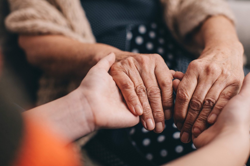
TREM2, Microglia, and Alzheimer’s: The Final Puzzle Piece?

       Alzheimer’s researchers have spent years focusing on the “amyloid hypothesis” proposing that toxic ꞵ-amyloid (Aꞵ) proteins, and the plaques they form outside neurons, were the primary cause of Alzheimer’s disease (AD). However, even therapies that successfully cleared Aꞵ in symptomatic individuals failed to slow or stop neurodegeneration, suggesting something else may be at play. Further research showed that Aꞵ buildup instigates the dysregulation of tau protein forming neurofibrillary tangles (NFTs) that aggregate inside neurons. When NFTs form near amyloid plaques the tau proteins may become detached, causing neuronal dysfunction. In this form, they are called neuritic plaques. On top of these complex processes, an intermediate step between Aꞵ build-up and the formation of neuritic plaques mediated by microglia may exist.

       Under normal circumstances, microglia are the brain’s immune cells and support neurons to combat pathogens and repair injured tissues by absorbing/breaking down debris, excess synapses, and pathogens. In the case of AD, one study found that microglia surround Aꞵ plaques and interact with the fibers, potentially preventing further aggregation or limiting the effects of toxic Aꞵ, supported by another study that found that fewer microglia around plaques predicted greater damage to nearby axons. Several AD risk-factor genes are expressed predominantly in microglia as opposed to neurons. For example, the R47H variant of the TREM2 gene, which codes for a microglial receptor, seems to impair microglial function and predicts more neuritic plaques and axonal damage. When the TREM2 gene was completely knocked out in mice with amyloid pathology microglia suffered major functional deficits, inhibiting them from interacting with amyloid plaques and leading to an increase in swollen, damaged neurons.

       If this R47H variant impairs microglial function thereby increasing neurodegeneration, then improving microglial function should be protective. Unfortunately, the answer is not so cut and dry. In mice with amyloid pathology but no tau pathology, this theory holds true and microglial activity seems to protect the brain. However, once tau pathology presents in mice, microglia modify their genetic expression activating genes that synergize with the presence of the APOE4 gene and are associated with greater neurodegeneration. To confirm this, researchers knocked out the TREM2 gene in mice with tau pathology and, unlike the previous knockout experiment with only amyloid pathology, this TREM2 knock out decreased neurodegeneration. In another study, mice with early tau pathology had their APOE4 levels reduced by half, resulting in decreased microglial activation and reduced neuronal damage.

       As these opposing results based on disease-phase show, the role of microglia in AD neurodegeneration (at least in mice) seems to change depending upon the presence or lack of tau pathology. To understand why this is, we must remember that microglia are immune cells that use cytokine signaling to communicate with other immune cells and mediate inflammatory responses. Research suggests that cytokines released by microglia exacerbates tau dysfunction and begin NFT aggregation. Evidence indicates that microglia have an important role in fighting amyloid buildup and pathology, protecting the brain in the early disease stages, but become detrimental once tau pathology begins as they overreact to the presence of neuritic (tau positive) plaques, increasing neurodegeneration and inflammation in response to the presence of misfolded tau.

       As such, researchers have begun to factor microglial activation per disease-stage into potential treatment models. Some suggest that it may be beneficial to up-regulate activation of microglia in the initial stages of amyloid build-up, allowing them to surround the plaques and prevent propagation of the pathology as long as possible. In fact, one such therapy is already being developed and tested for efficacy in clinical trials! AL002, developed by Alector, Inc., is a TREM2 agonist expected to upregulate microglial activity around amyloid plaques in the early disease stages prior to significant tau pathology, thereby slowing the rate of symptom onset and neurodegeneration. Excitingly, this trial will be using our facility as a research site so if you or someone you love is experiencing mild cognitive impairment or early Alzheimer’s disease, contact our office and we can discuss your eligibility for the trial. However, once tau pathology starts to develop, treatments would theoretically have to target inactivation of microglia in order to prevent their destructive effects on neurons via cytokine interaction with neuritic tau plaques, but no trials are yet testing this hypothesis.

Share on facebook
Facebook
Share on twitter
Twitter
Share on email
Email
Sources:
       Ulrich, J. & Holtzman, D.M. A New Understanding of Alzheimer’s [Magazine]. Scientific American. 2021.
       Gratuze, M., Leyns, C.E., & Holtzman, D.M. New insights into the role of TREM2 in Alzheimer’s disease [Online]. Molecular Neurodegeneration. 2018. Available from: https://molecularneurodegeneration.biomedcentral.com/articles/10.1186/s13024-018-0298-9.
       Mrak, R.E., Sheng, J.G., & Griffin, W.S. Glial cytokines in Alzheimer’s disease: review and pathogenic implications [Online]. Human Pathology. 1995. Available from: https://pubmed.ncbi.nlm.nih.gov/7635444/.

Donanemab Research Links Amyloid and p-tau217

       The FDA approval of Biogen’s aducanumab, now known as Aduhelm, set a low bar that other potential antibody treatments for Alzheimer’s disease are also hopeful to meet. Researchers from Eli Lilly recently reported that donanemab administered at plaque-dissolving strength correlated with diminishing levels of plasma p-tau217. When their data was plugged into a disease-progression model, it suggested that the decrease in amyloid and tau correlated with slower cognitive decline. Similarly, the Phase 2 trial and open-label extension of Biogen/Eisai’s lecanamab provided evidence of the drug’s disease modifying effect. The FDA has granted both donanemab and lecanamab breakthrough therapy status, and both Lilly and Biogen/Eisai have announced they will be using their Phase 2 data to seek accelerated approval.

       The most recent analysis of donanemab was carried out using data from Lilly’s TRAILBLAZER trial. During this trial, 131 participants randomized into the treatment group received infusions each month, with a stable dose of 1,400 mg after the first three half-doses at 700 mg. Once the participant’s amyloid burden fell below 25 centiloids, the dose was lowered to 700 mg. If the amyloid burden remained below 25 for two consecutive scans or if it fell below 11, participants were switched to placebo. By the end of the 76-week trial, results showed a 32 percent slowing of decline on the Integrated Alzheimer’s Disease Rating Scale (iADRS). Participants who cleared amyloid below 11 centiloids and were switched to placebo by 24 weeks showed amyloid burdens barely above 11 centiloids at 76 weeks. For this group, it would take 14 years for amyloid to accumulate back to baseline levels of approximately 90 centiloids. They also show reduced tangle burden in the temporal, parietal, and frontal lobes compared to participants in the placebo group.

       Several sources have reported that p-tau217 presence in CSF and plasma correlates with disease trajectory and amyloid and tangle burden. After 12 weeks of donanemab treatment, plasma p-tau217 fell significantly from subject-matched baseline measurements. This trend continued and strengthened in statistical significance during each follow-up assessment. At the end of the trial, the placebo group had a 6 percent increase in plasma p-tau217, while this level had decreased 24 percent from baseline in the treatment group. The drop in p-tau217 strongly correlated with a reduction in amyloid and tau tangles, suggesting that neurons release phospho-tau when amyloid is present. Thus, plasma p-tau217 may be used as biomarker to assess the effects of novel treatments on beta-amyloid fibrils. If its clinical efficacy is confirmed, it will be greatly useful in the development of Alzheimer’s treatments.

       Overall, the most intriguing implication of the donanemab data is that patients may only need to be treated for a few months to see lasting results. The link between amyloid and p-tau217 means the results could be easily and inexpensively tracked due to the simplicity of access to plasma, compared to current ways of testing amyloid levels with PET scans or spinal taps. Furthermore, this would be the strongest evidence that amyloid deposition induces tangle formation. If donanemab receives FDA approval, it may soon become a common treatment for seniors experiencing memory problems and even those preclinical patients, who are cognitively normal but at risk for developing Alzheimer’s disease. As Eli Lilly, Biogen, and Eisai begin the process of fast-track approval, we will likely know more in the coming months.

Share on facebook
Facebook
Share on twitter
Twitter
Share on email
Email
Source: 
On Donanemab, Plaques Plummet. Off Donanemab, They Stay Away. (2021, August 6). ALZFORUM. https://www.alzforum.org/news/conference-coverage/donanemab-plaques-plummet-donanemab-they-stay-away

The Case for the Amyloid Hypothesis

       The amyloid hypothesis has inspired most of Alzheimer’s research for the last 20 years. It proposes that accumulation of a protein fragment known as beta-amyloid, and in particular the 42 amino acid subtype, is the underlying cause of the Alzheimer’s pathogenesis. It is a “sticky” compound that builds up within the brain 10-15 years before symptoms emerge. In theory, it disrupts synaptic communication, and ultimately ends in neuronal death by initiating neural fibrillary tangle (NFT) formation. The theory posits overproduction, poor disposal, and eventual accumulation of beta-amyloid is the primary cause of Alzheimer’s disease. 

       Beta-amyloid is only a small section of the larger amyloid precursor protein (APP). At present, scientists have not determined the normal function of APP. However, they know that when the protein is activated, it is divided into smaller pieces that are both inside or outside of cells. When pieces of beta-amyloid begin to accumulate, they form small clusters called oligomers. Multiple recent studies have implicated beta-amyloid oligomers as upstream drivers of Alzheimer’s disease. As these oligomers accumulate in the brain, they create chains of clusters known as fibrils, which then bond together to form beta-sheets. The characteristic amyloid plaques found in Alzheimer’s patients are composed of beta-sheets meshed together with other substances, such as tau protein making up NFTs. These plaques are believed to create intra-neuronal NFTs that cause synaptic injury, loss of neuronal function, and eventually cell death.

       Genetic studies of familial Alzheimer’s patients identified over 200 mutations that lead to an early onset of the disease. Many of these mutations occur in the APP gene and cause an increase in the production of beta-amyloid. Overproduction causes an increase in misfolded beta-amyloid, resulting in the formation of amyloid oligomers and eventually plaques.

       The APOE4 genotype has been identified as the greatest risk factor for developing Alzheimer’s disease. Those who carry the E4 allele of apolipoprotein E (APOE) have a much greater risk for developing early onset Alzheimer’s disease. The APOE4 genotype, which has been identified in approximately 65% of Alzheimer’s patients, is associated with reduced ability to clear amyloid from the brain and increased aggregation of beta-amyloid into oligomers. 

     Another argument for the amyloid hypothesis comes from examining the brains of individuals with Down syndrome. Adults with Down syndrome have three copies of the chromosome carrying the APP gene: chromosome 21. These individuals invariably develop beta-amyloid plaques identical to those seen in Alzheimer’s disease and have a high risk of developing early onset dementia. Thus, overproduction of beta-amyloid plaques in Down syndrome results from a comparable genetic pathology to Alzheimer’s patients with mutations of the APP gene.

       Clinical trials of anti-amyloid agents that target beta-amyloid oligomers, such as aducanumab, donanemab, and lecanemab, have exhibited statistically significant clinical efficacy. Though as discussed in previous blog posts, the benefit received from these drugs is directly related to how early they’re given in the progression of the disease, generally before amyloid plaques have accumulated to the point of causing too much neural damage, which suggests that beta-amyloid accumulation initiates the cascade of Alzheimer’s pathogenesis. 

       The genetic and clinical trial data suggests beta-amyloid plays a key part in Alzheimer’s disease. The research sparked by the amyloid hypothesis remains relevant to this day and continues to inspire further clinical investigation. However, there is further emerging evidence that suggests it is far from the only factor influencing pathogenesis. Future studies targeting beta-amyloid and further examining APP mutations may one day lead to promising therapeutic interventions for Alzheimer’s disease, but alternative methods should also be pursued. Check in next week when we’ll be discussing these alternative methods.

Share on facebook
Facebook
Share on twitter
Twitter
Share on email
Email
Sources:
Annus, T., Wilson, L. R., Hong, Y. T., Acosta–Cabronero, J., Fryer, T. D., Cardenas–Blanco, A., Smith, R., Boros, I., Coles, J. P., Aigbirhio, F. I., Menon, D. K., Zaman, S. H., Nestor, P. J., & Holland, A. J. (2016). The pattern of amyloid accumulation in the brains of adults with Down syndrome. Alzheimer’s & Dementia, 12(5), 538–545. https://doi.org/10.1016/j.jalz.2015.07.490
Beta-amyloid and the Amyloid Hypothesis. https://www.alz.org/national/documents/topicsheet_betaamyloid.pdf
Tolar, M., Hey, J., Power, A., & Abushakra, S. (2021). Neurotoxic Soluble Amyloid Oligomers Drive Alzheimer’s Pathogenesis and Represent a Clinically Validated Target for Slowing Disease Progression. International Journal of Molecular Sciences, 22, 6355. https://doi.org/10.3390/ijms22126355


Why has Biogen’s aducanumab become so controversial?

The FDA’s approval of aducanumab has been shrouded by controversy, with experts quickly criticizing the FDA’s ruling, referencing two large studies showing little convincing evidence of efficacy. These feelings were mirrored by the FDA’s own advisory committee. Of the 11 members, 10 voted against the drug’s approval, citing insufficient evidence, and one member was undecided. The drug was approved anyway, and three committee members have since resigned.

To understand the reasoning behind their actions, let’s discuss the science behind the drug’s approval. Biogen’s aducanumab, now marketed under the name Aduhelm, is an amyloid antibody that works to clear β-amyloid from the brain. Previous drugs have successfully cleared amyloid from the brain but did not improve cognitive function. Most of Biogen’s trials produced similar results.

Only after re-analysis was Biogen able to show cognitive decline slowed in a subset of patients from their EMERGE trial. The positive effects were seen only in patients who carried the APOE4 genotype, while patients who were not carriers fared worse on the drug than the placebo. The identical ENGAGE trial failed to produce positive results altogether.

The negative results from the ENGAGE trial are important because the positive effects seen in APOE4 patients from the EMERGE trial only began to occur following a change in study protocol. Initially, APOE4 patients were receiving lower doses of the drug because they are more prone to amyloid-related imaging abnormalities (ARIA) while receiving higher doses. Midway through the trial, this was adjusted so that all patients were receiving the same dose per kilogram. This caused more APOE4 patients to experience imaging abnormalities, making it necessary to pause dosing and MRI monitoring until the condition cleared. Therefore, many of these patients and their physicians became aware that they were likely receiving the drug, breaking the blind and possibly resulting in bias data.

Biogen maintains that the increased dosage produced greater efficacy in this subset of patients following the change in protocol and used this data to obtain FDA approval. However, positive results may have stemmed from the unintentional unblinding of the treatment group, potentially affecting ratings on measures of cognitive function and daily living. Thus, the drug was approved based on potentially erroneous data. 

An independent analysis of the heterogeneity of Alzheimer’s disease progression points to further issues with the EMERGE and ENGAGE results. When comparing individual trajectories on cognitive assessments, performance was highly variable in prodromal and mild Alzheimer’s disease patients. When these results were compared to the data from actual clinical trials (i.e. Biogen’s EMERGE and ENGAGE), the differences fell within the range expected when there is no treatment effect. Therefore, it’s also important to consider that the results generated by Biogen could be due to oversampling of individuals who are declining faster in the placebo group or declining slower in the treatment group.

Following aducanumab’s approval, it is unclear if it will become the new standard that potential new treatments are compared to instead of placebo. What we do know now is that Biogen will be required to conduct a new randomized controlled phase 4 trial to verify the efficacy of aducanumab. During the first two years, Biogen will have a monopoly on the market and participants will be required to pay approximately $56,000 upfront as it stands now, although some supplemental insurances may reduce this rate. Even if Medicare does fully reimburse, the burden on the system would be significantly higher than any other drug currently on the market. If they decide not to cover the cost, very few people will be able to utilize it.

Moreover, the drug relies heavily on the hypothesis that amyloid is the underlying cause of Alzheimer’s disease. If a patient has received an amyloid PET scan and knows their APOE status, then the drug may be worth it in the early stages of the disease. However, if the disease has progressed too far, removing amyloid doesn’t translate to improved cognition or functioning.

All in all, whether aducanumab is worth it depends on the patient and the situation. If you’re in the early stages of the disease, know your APOE status, have had an amyloid PET scan, and are willing to pay upfront for treatment, it may be worth your while. However, few people fall into this category. Our hope is that research will continue to explore other options independently and without comparison to Biogen’s aducanumab.

Share on facebook
Facebook
Share on twitter
Twitter
Share on email
Email
Sources:
https://n.neurology.org/content/96/22/e2673
https://time.com/6072980/alzheimers-drug-approval-controversy/
https://www.alzforum.org/news/research-news/aducanumab-still-needs-prove-itself-researchers-say#enlarge
https://www.kff.org/medicare/issue-brief/fdas-approval-of-biogens-new-alzheimers-drug-has-huge-cost-implications-for-medicare-and-beneficiarie

Donanemab: A Promising Anti-Amyloid Therapy

     Due to inconsistent effects of anti-amyloid therapies, such as aducanamab, a recent shift from the “amyloid hypothesis” to the “tau hypothesis”, has occurred. The primary issue with most anti-amyloid therapies, is that the successful removal of amyloid-β (Aβ) plaques doesn’t significantly reduce the clinical symptoms, suggesting that significant damage to the underlying brain structures has already occurred. However, donanemab, another anti-amyloid therapy, promises to be different, and here’s how.

        Firstly, donanemab, unlike most of the previous anti-Aβ therapies, is designed for early use in Alzheimer’s disease (AD), when Aβ levels are notable yet phosphorylated tau (pTau) making up neurofibrillary tangles (NFTs) are not widely deposited. The presence of amyloid is believed to form pTau misfolding and aggregation into NFTs. Not including individuals with widely dispersed NFTs helped provide a clearer picture of what Aβ removal can improve, without significant NFT presence contributing to continued dysfunction and cell death. Secondly, in a Phase-II trial donanemab met its primary outcome measure, producing a significant improvement in 2 cognitive/functional rating scales compared to controls. The secondary outcomes did not reach significance in this trial, but likely could have in a trial with more participants (such as the stage 3 trial with donanemab that our site is participating in). Finally, it targets a specific type of Aβ known as N3 truncated, pyroglutamylated Aβ (pE-Aβ) which is present in relatively low quantities compared to other types of Aβ but is especially prone to “seeding” or propagation of Aβ pathology by interacting with and misfolding other proteins.

      This pE-Aβ has been researched previously and is strongly implicated in the pathogenesis of AD. Its role in neurodegeneration was confirmed in AD mouse when its inhibition improved memory and decreased Aβ deposition. This is because pE-Aβ works synergistically with conventional Aβ when co-incubated, forming resilient hybrid oligomers that have stronger cytotoxic effects than oligomers formed by non-pE-Aβ alone. Therefore, a drug such as donanemab that targets pE-Aβ before they initiate the cascade of plaque formation could, in theory, prevent significant plaque-induced neurodegeneration.

         Even more exciting, in the Phase-II trial also improved Aβ clearance with spectacular results such that within 24 weeks of starting treatment, 25% of the subjects on active drug reached Aβ negativity as reflected in Amyloid-PET scans. By week 76, 68% were Aβ negative. Furthermore, as expected, these results were strongest in those with lower pTau burden at baseline. On the other hand, there were also unexpected outcomes such as the lack of significant hippocampal volume change and an overall decrease in brain volume and increase in ventricular size for those in the treatment group compared to controls. Researchers hypothesized this was due to the rapid removal of plaque volume. Despite these promising outcomes, donanemab is not expected to be a sole treatment for AD in the future. Alzheimer’s is a multi-factorial disease and therefore researchers expect to someday utilize a multi-factorial combination of therapies to specifically target each phase of the disease spectrum and the new Phase-III TRAILBLAZER-ALZ2 trial for donanemab may emerge as one of these future treatments!

Share on facebook
Facebook
Share on twitter
Twitter
Share on email
Email
Sources:
Talan, J. A New Monoclonal Antibody Shows Promise Early in Alzheimer’s Disease [Article]. Neurology. 2021.
Nussbaum, J., et al. Prion-like behavior and tau-dependent cytotoxicity of pyroglutamylated amyloid-β [Online]. Nature. 2012. Available from:  https://www.ncbi.nlm.nih.gov/pmc/articles/PMC3367389/

Lion’s Mane: A Mushroom to Remember

       This week we will be discussing the mushroom Lions Mane, or Hericium Erinaceus, and its health applications for Alzheimer’s Disease (AD). Currently, there are no drugs on the market that prevent, reverse, or halt AD progression. Although a few clinical trials in the pipeline show promise, scientists are also looking to alternative treatments, like Lions Mane, to combat AD.

       Lions Mane is a culinary mushroom and is commonly eaten in countries such as Japan and China without any harmful effects. It’s generally found growing under old broadleaf trees and contains erinacines, natural substances with potential pharmacological effects on the central nervous system (CNS). There are two forms of Lions Mane that can be ingested; the fruiting body and the mycelium that encompasses the erinacines. Erinacines fall within a group of compounds called cyathin diterpenoids, and are stimulators of nerve growth factors (NGF). Nerve growth factors play a supportive role in the CNS, and are critical to adequately protect surviving and developing neurons.

       Rats given erinacine A for 3 weeks increased their concentrations of noradrenaline and homovanillic acid in the hippocampus compared to controls and showed evidence of an overall increase in NGF levels, most notably in the dentate gyrus of the hippocampus. Increases in noradrenaline and homovanillic acid result in more alertness and better retrieval of memory along with breaking down fat and increasing blood sugar levels to promote more energy, suggesting that erinaceus A promotes nerve and brain health in animal models. Furthermore, H. erinaceus mycelium containing erinacine A administered orally to AD transgenic mice for 30 days resulted in decreased recruitment and activation of plaque burden compared to controls. Other structures often impaired in AD, like the hippocampus and the locus coerulous, also showed functional improvement compared to those not given the mycelium.

       A double-blind clinical trial assessing the oral administration of H. erinaceus fruiting bodies in elderly humans showed improvement in subjects with mild cognitive impairment compared to age-matched controls. Researchers measured improvements using the Revised Hasegawa Dementia Scale (HDS-R). The group ingesting H. erinaceus significantly increased their scores during the 16-week treatment period, indicating improvement compared to those not taking H. erinaceus. However, when subjects stopped taking H. erinaceus their scores began to fall, reflecting scores similar to those that were untreated, indicating the need for continued use.

       Several different compounds in H. erinaceus appear to contain protective benefits, such as amyloid plaque reduction, insulin-degrading enzyme expression, enhancing NGF release, and even managing neuropathic pain. Although some is known about erinacines, many of the compounds are still undergoing research, with some still being discovered. These discoveries and continued ones will hopefully continue to pave the way for therapeutic strategies to prevent, manage, and slow AD progression. Furthermore, prior research and clinical trials have proven that Lion’s Mane and its extracts are safe for human consumption at doses of 3-4 grams per day (although allergies have been noted). If this research interests you, we encourage you to discuss Lion’s Mane with your primary care physician to potentially improve your cognition.

Share on facebook
Facebook
Share on twitter
Twitter
Share on email
Email
Sources:
Li, I. C., et al. NeuroHealth Properties of Hericium erinaceus Mycelia Enriched with Erinacines [Online]. 2018. Available from: https://pubmed.ncbi.nlm.nih.gov/29951133/
Mori, K., et al. Nerve Growth Factor-Inducing Activity of Hericium erinaceus in 1321N1 Human Astrocytoma Cells [Online]. 2008. Available from: https://pubmed.ncbi.nlm.nih.gov/18758067/
Chong, P. S., Fung, M. L., Wong, K. H., & Lim, L. W. Therapeutic Potential of Hericium erinaceus for Depressive Disorder [Online]. 2019. Available from: https://pubmed.ncbi.nlm.nih.gov/31881712/
Mori, K., Inatomi, S., Ouchi, Y., Azumi, Y., Tuchida, T. Improving effects of the mushroom Yambushitake (Hericium erinaceus) on mild cognitive impairment: a double-blind placebo-controlled clinical trial [Online]. 2009. Available from: https://pubmed.ncbi.nlm.nih.gov/18844328/
Shimbo, M., Kawagishi, H., Yokogoshi, H. Erinacine A increases catecholamine and nerve growth factor content in the central nervous system of rats [Online]. 2005. Available from: https://www.sciencedirect.com/science/article/abs/pii/S0271531705001041
Chen, C., et al. Erinacine S, a rare sestererpene from the mycelia of Hericium erinaceus [Online]. 2016. Available from: https://pubmed.ncbi.nlm.nih.gov/26807743/

The Curious Case of Alzheimer’s-Related Primary Progressive Aphasia

This week we review a disease called Alzheimer-related primary progressive aphasia (PPA-AD). It is well known that a primary symptom of Alzheimer’s disease (AD) is memory impairment, while the primary symptom of primary progressive aphasia (PPA) is an isolated language disturbance. Two thirds of PPA cases are caused by a tauopathy called Frontotempral degeneration (FTD) but the remaining third are due to AD pathology making up the PPA-AD group.

            The fact that PPA-AD has only language dysfunction as opposed to memory impairment has perplexed researchers for years. Recently, a group found a possible explanation for this during a longitudinal follow-up on a cohort (n = 31) with AD, divided into typical amnestic presentations (n = 14) versus language presentation (PPA-AD, n = 17). Participants received longitudinal memory and language tests, biomarker analyses, and the majority agreed to autopsy. Using these metrics, they discovered some trends that may shed light on why PPA-AD and typical AD dementia (DAT-AD) have such different clinical presentations.

            In the PPA-AD group, participants had a significant yearly decline of 6.21% in object naming scores and 4.25% for a measure of global language performance, with no significant decrease in memory. Those in the DAT-AD group, meanwhile, had a significant yearly decrease of 2.15% for memory and 4.05% for object naming. Structural imaging was also done on the PPA-AD group at their initial visits showing cortical thinning, especially of the language-dominant left hemisphere, extending throughout the language network. Significant thinning of the parahippocampal gyrus was present only on the left side (as shown below, denoted by PHG).

To assess neuropathology, Aβ plaques, neurofibrillary tangles (NFTs), and overall plaque density were quantified. The PPA-AD group showed maximum levels of Aβ and NFTs, though researchers focused on NFTs as their distribution and density more strongly correlates to cognition. Specifically, the PPA-AD group had severe NFT pathology in the neocortex and all medial-temporal lobe structures associated with memory. However, 2 of the 8 who agreed to undergo autopsy were found to be of the “hippocampal-sparing type” where cortical NFT density is higher than in memory-related structures. Interestingly, despite the decrease in hippocampal NFT density and sparing of memory, those 2 participants had severe NFT-induced degeneration of memory regions. Furthermore, bilateral comparisons revealed that the left hemisphere of these 2 participants had more Aβ plaques while other patients had elevated NFTs, suggesting a dichotomy between pathogenesis of hippocampal-sparing type PPA-AD and typical PPA-AD.

            This leaves the question, what induces resilience of memory structures in PPA-AD? Well, in PPA-AD the hippocampal gyrus primarily degenerates on the left hemisphere. Previous lesion studies on this area have shown that episodic memory function only significantly declines when bilateral lesioning or degeneration of hippocampal structures occurs. However, while this study showed decreased NFT aggregation in memory-related structures in PPA-AD compared to cortical areas, previous studies have had varied results indicating that further longitudinal studies of this type are required.

One other possible explanation for memory resilience in PPA-AD involves APOE status. The PPA-AD group had a 14.7% incidence of ε4 alleles, which inhibits neuronal plasticity, while the rest had ε3 alleles, which enhance neuronal plasticity. Interestingly, this frequency of ε4 alleles matches control populations with PPA-AD, while the DAT-AD group had a 42% ε4 frequency with 3 homozygous carriers, almost twice as high as control populations. Thus ε4 presence likely increased vulnerability of memory networks in the DAT-AD group by inhibiting compensatory mechanisms like neuroplasticity, while those with PPA-AD, having reduced ε4 and increased ε3 frequency, may increase resilience to neurodegeneration by allowing for neuronal plasticity.

Share on facebook
Facebook
Share on twitter
Twitter
Share on email
Email
Sources:
Sajjadi, S. A., Ash, S., & Cappa, S. Preservation of Memory in Alzheimer’s-Related Primary Progressive Aphasia [Magazine]. Neurology. 2020.
Preib, D., Billette, O. V., Schneider, A., Spotorno, N., & Nestor, P. J. The atrophy pattern in Alzheimer-related PPA is more widespread than that of the frontotemporal lobar degeneration associated variants [Online]. NeuroImage: Clinical. 2019. Available from: https://www.ncbi.nlm.nih.gov/pmc/articles/PMC6734177/
Mesulam, M. M., et al. Memory Resilience in Alzheimer Disease With Primary Progressive Aphasia [Online]. Neurology. 2021. Available from: https://n.neurology.org/content/96/6/e916.long
Primary Progressive Aphasia [Online]. National Aphasia Association. Available from: https://www.aphasia.org/aphasia-resources/primary-progressive-aphasia/

How PWAS Helped Discover 10 New Alzheimer’s Genes

       For quite a while, one of the most common methods of isolating disease-risk-associated genes has been a Genome Wide Association Study (GWAS) which involves genotyping a large number of people and associating various genetic loci, or locations, with the phenotypes, or visible traits, that commonly arise from variance at these loci. However, researchers recently developed an even more informative way of performing these genomic analyses, using it to discover 10 new genes that may modify risk of Alzheimer’s disease (AD)! This new method is very similar to GWAS but adds another layer of data regarding protein function, becoming a Proteome-Wide Association Study (PWAS).

       PWAS expands upon the information provided by GWAS by analyzing the type of mutation present and quantifying the change in production or functionality of the protein produced by that gene in comparison to controls. Next, protein function scores are correlated to the phenotypes of subjects with those genes to confirm the expected effects. In analysis of a binary phenotype (a trait that is either present or absent with no intermediate presentation) a strong correlation is derived when subjects with a disorder have a significantly different functional effect score than controls, confirming PWAS’s prediction that the protein is less (or more) functional in the mutated form.

Figure 1. A diagram depicting the difference in analyses between Genome-Wide Association Studies and Proteome-Wide Association Studies.

       The additional information provided by PWAS allows detection of associations that are not detectable by GWAS. Researchers at Emory University recently took advantage of this, using PWAS to discover 10 new genes associated with AD risk. To begin, they isolated 1,475 genes whose abundance is genetically controlled and analyzed their AD-risk score in a GWAS dataset (71,880 cases, 383,378 controls). Of the 1,475 genes, only 13 were related to AD risk in the GWAS dataset.

       After further analyses with PWAS, including causality tests and adjustments for APOE status, 11 genes remained with evidence for causality of AD. Only 1 of these genes, ACE, had previously been strongly correlated with AD but the other 10 are relatively new discoveries! Those genes include syntaxin 4 and 6, DOC2A, SNX32, ICA1L, cathepsin H, CARHSP1, LACTB, RTFDC1, and PLEKHA1. Although little is known about their relationship to AD pathogenesis, researchers will undoubtedly begin researching how these genes relate to AD risk and development. In time you may even see treatments emerging targeting these genes or proteins.

       Even more interestingly, several of these genes impact molecular pathways that are not widely considered part of the disease model for AD, such as LACTB which is a mitochondrial protein (with mitochondrial dysfunction only getting attention as a possible mechanism of AD development within the last few years) and PLEKHA1 which mediates transmembrane signaling more generally. This suggests that there are as-of-yet undiscovered or under-researched factors that relate to AD risk and pathogenesis, opening the door for new treatments and a more complete understanding of the disease itself.

Share on facebook
Facebook
Share on twitter
Twitter
Share on email
Email
Sources:
PWAS x GWAS? Proteome Analysis Nets 10 New Alzheimer’s Genes [Internet]. Alzforum. 2021. Available from: https://www.alzforum.org/news/research-news/pwas-x-gwas-proteome-analysis-nets-10-new-alzheimers-genes
Brandes, N., Linial, N, & Linial, M. PWAS: Proteome-Wide Association Study [Internet]. bioRxiv. 2019. Available from: https://www.biorxiv.org/content/10.1101/812289v1.full

Pollution and Plaques: How Your Environment Impacts Neurodegeneration

       It was previously thought that fine particulate matter, produced by car exhaust and wildfires among other things, makes its way into the brain where it damages neurons and glia directly or damages brain vasculature leading to vascular dementia. However, young people in Mexico City (a high pollution area) have also been shown to already possess amyloid plaques, tau neurofibrillary tangles, and alpha-synuclein aggregates in the brain suggesting that pollution may also lead to dementia via non-vascular mechanisms. Two more recent studies, led by Gil Rabinovici at UC San Francisco and Diana Younan at USC Los Angeles, decided to expand on this research.

       Gil and his colleagues used the IDEAS cohort PET scans and related the amyloid burden to air pollution levels in the ZIP codes of participants 1-2 years and 13-15 years before their scan. Those who lived in areas of higher pollution (measured in µg/m3 or PM2.5) were 1.15 times more likely to have a positive amyloid PET per 4 µg/m3 increase. Melinda Power of George Washington University wants to expand this study further to include measures of residential history and occupational pollution exposure, warning that ‘Area-level exposure’, such as that of an entire ZIP code, ‘may or may not be representative of individual-level exposure’.

       Meanwhile, Diana Younan and colleagues looked for a relationship between air pollution and brain atrophy of AD-associated structures in cognitively normal older women. They used MRIs from the WHIMS cohort (712 women who received an MRI scan in 2005-2006 and again in 2010-2011) and their addresses (taken twice a year). They observed that for every 2.82 µg/m3 increase in PM2.5, participants experienced gray matter atrophy associated with a 24% increase in AD risk! What is even more concerning is that these findings, and Gil’s, came from participants who, largely, live in areas that are within the Environmental Protection Agency’s acceptable pollution levels. This means that toxin levels that the EPA determines to be “safe” are already increasing the risk of cognitive impairment, highlighting the need for cleaner/greener ways of living.

       Overall, these studies link pollution to dementia widely and to AD dementia in the form of amyloid accumulation and brain atrophy. The fact that the results remain consistent despite different cohorts and different testing paradigms is compelling but epidemiologists want to see further replication, stating that the direction of the relationship between air pollution and specific neuropathologies is not clear enough yet. Luckily, air quality in the US has improved steadily since 1990 when the Clean Air Act was amended but, as these studies show, these patterns exist even at the currently accepted levels of air quality, leaving room for improvement.

Share on facebook
Facebook
Share on twitter
Twitter
Share on email
Email
Sources:
You Are What You Breathe: Polluted Air Tied to Plaques, Brain Atrophy [Internet]. 2020. Alzforum. Available from: https://www.alzforum.org/news/research-news/you-are-what-you-breathe-polluted-air-tied-plaques-brain-atrophy.
Iaccarino, L., et al. Association Between Ambient Air Pollution and Amyloid Positron Emission Tomography Positivity in Older Adults With Cognitive Decline [Internet]. JAMA Neurology. 2020. Available from: https://pubmed.ncbi.nlm.nih.gov/33252608/.
Younan, D., et al. PM2.5 associated with gray matter atrophy reflecting increased Alzheimers risk in older women [Internet]. Neurology. 2020. Available from: https://pubmed.ncbi.nlm.nih.gov/33208540/.

Further Breakthroughs in AD Testing

       A couple months ago we posted a blog about a blood test for Alzheimer’s disease (AD) that changed the game of diagnosing AD. Unsurprisingly, the scientific community immediately began researching and attempting to improve the test and already they have made significant progress! This week we will break down what we have learned about the AD blood test, why it matters, and what remains to be seen.

       Amyloid-β (Aβ) residues and various sub-types of the AD biomarker protein tau are present both in the blood and in the brain. The basis of the AD blood test involves identifying how much of the Aβ and tau sub-types (primarily p-tau181 and p-tau217) are present in serum and correlating these levels to those in the brain to assess increased risk of AD. The initial findings promised a test to identify AD biomarkers, and therefore risk of developing AD, long before symptoms begin.

       Continued research supported the accuracy of the tests ability to identify AD biomarkers years before PET imaging and decades before onset of symptoms. The blood test was recently approved under the Clinical Laboratory Improvements Amendment protocol meaning that it is an accepted method of testing human samples outside of research settings.

       Researchers continued to compare the accuracy of results of p-tau181 levels, Aβ42/40 ratios, and neurofilament light (NfL) testing to see which may be a better predictor of assessing AD risk. P-tau181 levels combined with NfL testing proved the best method. Together, they can differentiate between AD and other dementing illnesses such as Frontotemporal Dementia and Progressive Supranuclear Palsy with 88% accuracy!

       On top of its use for diagnostic purposes, this testing method can also predict disease progression. One study showed that groups with higher p-tau181 levels progressed more quickly over the course of 2.5 years compared to those with lower p-tau181 levels. Other studies suggest this trend may even apply at the individual level with testing of p-tau181 and NfL predicting progression to AD over the course of 4 years in two separate studies of 107 and 74 participants respectively. Once again, the predictions were 88% accurate!

       However, despite all of this promise, some experts in the field aren’t convinced and would like to see more results. Oskar Hansson of Lund University says that he believes tau serum testing is a great method for diagnosing AD in those with MCI/dementia but that diagnosing preclinical AD will require a combination test for multiple blood-based markers. Furthermore, while p-tau181 tests have been quite useful, p-tau217 is seemingly even more useful with one study suggesting it might have twice the predictive power in comparison to p-tau181. As you can see, tau serum blood tests have opened up a new door into the progression of AD but studies remain to be done on how to most effectively use this new tool.

Share on facebook
Facebook
Share on twitter
Twitter
Share on email
Email
Source:
Plasma Aβ Test Wins Approval – Are p-Tau Tests Far Behind? [Internet]. 2020. Alzforum. Available from: https://www.alzforum.org/news/research-news/plasma-av-test-wins-approval-are-p-tau-tests-far-behind