The Gut-Brain Axis: Part 2 of 3

       As we discussed last week, our microbiome has profound impacts outside of the digestive system including the central nervous system (CNS). The interaction within the Gut-Brain Axis is reciprocal; our brain communicates with our gut microbes via the enteric nervous system (ENS) consisting of over 100 million neurons responsible for modulation of intestinal contraction patterns, secretion of enzymes, and regulation of blood flow necessary for digestion. Through regulation (or dysregulation) of these functions the brain has a direct impact on the microbiome. Mice exposed to 1 hour of psychological stress for a week have irregular concentrations of microbes associated with intestinal disease. 

       When compared to mice not exposed to stress, the greatest change in microbial concentration was the loss of the bacteria Akkermansia muciniphila (roughly 1-4% of total microbiota) this loss was associated with increased insulin resistance, inflammatory responses, and metabolic endotoxemia.  Brain insulin receptors are concentrated in the hippocampus, a key structure for memory function.

       Insulin resistance is associated with an increased risk for Alzheimer’s disease (AD). Healthy insulin signaling aids in the reduction of neuroinflammatory responses in the ENS and brain. Thus, stress induces an unhealthy feed-back loop in the microbiome that acts back on the brain to cause insulin resistance, increased neuroinflammation and cognitive worsening.

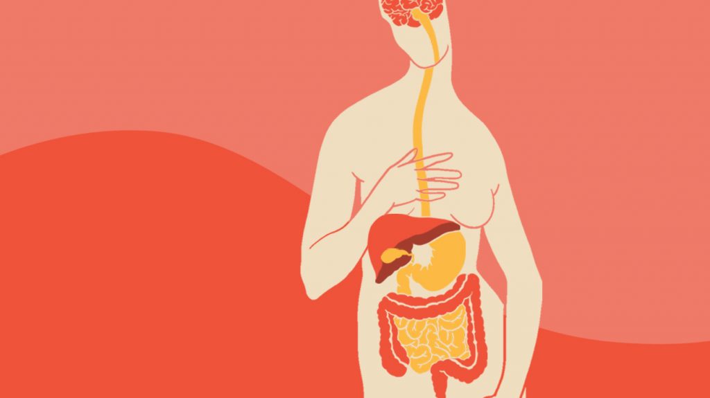
     This feedback loop between the brain and microbiome highlights the importance of maintaining a balance of both mental health and gut health. Communication between the brain and gut biome occurs through three pathways in the nervous, endocrine, and immune systems. These pathways regulate gut motility, gut permeability, and intestinal hormone secretions that effect microbial gene expression.

        The first of these mechanisms – gut motility, is regulated, but not initiated, by innervation from the cranial vagus nerve to the ENS in the form of the migrating motor complex (MMC). The MMC is a distinct electromechanical pattern in intestinal smooth muscles present during fasting. Impaired MMC regularity, caused by disrupted sleep and mood, decreases intestinal movement allowing overgrowth of small bowel bacteria and lowering diversity in the distal gut.

     Second, the permeability of the intestinal barrier can be changed by stress causing a dysfunctional state known as “leaky gut” via changing epithelial cell permeability and the mucosal layer. In rats both acute and chronic stress increased leakiness of gut epithelial cells allowing microbes and their by-products into surrounding cells and the bloodstream where they caused inflammation all over the body. A “leaky gut” with an inflammatory immune response was also observed in mice after premature maternal separation and was reversed with antidepressant treatment. This result highlights the powerful role that the brain can have on the microbiome, and vice versa.

      Third, stress decreases secretion of mucus by goblet cells in the gut resulting in a less protective mucus layer increasing ulcer risk and impacting the microbiome, particularly A. muciniphila that lives in and feeds upon intestinal mucus metabolizing it into short chain fatty acids (SCFAs). These fatty acids aid glucose homeostasis, lipid metabolism, appetite regulation, serotonin synthesis, and even immune function. In a healthy gut A. muciniphila stimulates mucus production leading to a healthy gut, brain, and body.

      The microbes in our gut have binding sites for many of the molecules that our nervous, endocrine, and immune systems use to communicate. One example of this is serotonin, which can be released by enterochromaffin cells (ECCs) into the intestinal lumen where it acts upon Clostridiales, another gut microbe. In response to serotonin release, Clostridiales produce secondary bile acids and SCFAs that signal back to the ECC to upregulate serotonin signaling to the CNS through vagal/spinal circuits effecting mood regulation. Release of the “fight-or-flight” transmitter norepinephrine, increases virulent traits in microbes, as well as stimulating the growth of other strains of enteric pathogens. This may explain why strenuous life events that increase norepinephrine are associated with gastroenteritis and irritable bowel syndrome.

       It is clear that there are numerous interactions occurring bidirectionally within the gut- brain axis that impact the brain and the microbiome. Unfortunately, the interactions from the brain to the gut are not nearly as well studied as those in the other direction requiring further research to parse the specific ways that psychological distress might translate to intestinal dysfunction. Furthermore, there are several disorders, including AD, anxiety, depression, Parkinson’s, and even Autism Spectrum Disorder,  that may effect interactions in the gut-brain axis but very few studies are able to confirm the direction of causality, and even fewer studies do so in humans.  There is research on ways in which we can nurture a healthy gut microbiome for both gut and brain health
that we will address in the next blog. Join us next time for the final piece of the puzzle in part three of The Gut-Brain Axis!

Facebook
Twitter
Email
The Brain-Gut Connection [Internet]. Johns Hopkins Medicine. 2019. Available from:
https://www.hopkinsmedicine.org/health/wellness-and-prevention/the-brain-gut-connection
Martin, C. R., Osadchiy, V., Kalani, A., & Mayer, E. A. The Brain-Gut-Microbiome Axis
[Internet]. Cellular and Molecular Gastroenterology and Hepatology. 2018. Available from:
https://www.ncbi.nlm.nih.gov/pmc/articles/PMC6047317/
Aguilera, M., Vergara, P., & Martinez, V. Stress and antibiotics alter luminal and wall-adhered microbiota and enhance the local expression of visceral sensory-related systems in mice [Internet]. Neurogastroenterology & Motility. 2013. Available at:
https://onlinelibrary.wiley.com/doi/full/10.1111/nmo.12154
Mayer, E. A., Tillisch, K., & Gupta, A. Gut/brain axis and the microbiota [Internet]. The Journal of Clinical Investigation. 2015. Available at: https://www.jci.org/articles/view/76304
Noble, E. E., Hsu, T. M., & Kanoski, S. E. Gut to Brain Dysbiosis: Mechanisms Linking
Western Diet Consumption, the Microbiome, and Cognitive Impairment [Internet]. Frontiers in
Behavioral Neuroscience. 2017. Available at: https://www.ncbi.nlm.nih.gov/pmc/articles/PMC5277010/
Fujio-Vejar, S., Vasquez, Y., Morales, P., Magne, F., Vera-Wolf, P., Ugalde, J. A., Navarrete, P.,
& Gotteland, M. The Gut Microbiota of Healthy Chilean Subjects Reveals a High Abundance of the Phylum Verrucomicrobia [Internet]. Frontiers in Microbiology. 2017. Available at:
https://www.ncbi.nlm.nih.gov/pmc/articles/PMC5491548/
Tanaka, T., Kendrick, M. L., Zyromski, N. J., Meile, T., & Sarr, M. G. Vagal innervation
modulates motor pattern but not initiation of canine gastric migrating motor complex [Internet].
Neuroregulation and Motility. 2001. Available at:
https://www.physiology.org/doi/full/10.1152/ajpgi.2001.281.1.G283?url_ver=Z39.88-
2003&rfr_id=ori:rid:crossref.org&rfr_dat=cr_pub%3dpubmed
Morrison, D. J., & Preston, T. Formation of short chain fatty acids by the gut microbiota and
their impact on human metabolism [Internet]. Gut Microbes. 2016. Available at:
https://www.ncbi.nlm.nih.gov/pmc/articles/PMC4939913/
Rhee, S. H., Pothoulakis, C., Mayer, E. A. Principles and clinical implications of the brain-gut-
enteric microbiota axis [Internet]. Nature Reviews Gastroenterology & Hepatology. 2009.
Available at: https://www.ncbi.nlm.nih.gov/pmc/articles/PMC3817714/

The Gut-Brain Axis: Part 1 of 3

         We each have a unique and complex network of microbes living within our guts known as a microbiome. It is comprised of a dynamic ecosystem of viruses, fungi, and predominately bacteria. As humans, we have been evolving and diversifying in concert with our microbiomes for at least 15 million years, indicating a close bacteria-host mutualism. Colonization of your gut microbiome occurs during birth, is highly dynamic throughout infancy, and resembles adult microbiomes in approximately three years remaining stable thereafter. Several factors influence the microbiome, such as genetics, diet, metabolism, age, geography, stress, antibiotic treatment, probiotics, disease, and more. Microbes within the microbiome are responsible for extracting energy from the food we eat, vitamin biosynthesis, pathogen overgrowth protection, educating our immune system, and more.

     Alterations in the gut microbiome are associated with an array of metabolic and gastrointestinal diseases like irritable bowel syndrome (IBS) and insulin resistance, but recent research has found that other body regions are also affected. Your gut microbiome is important and key in regulating digestion, hormones, immunity, the brain and cognition. Due to this newly established relationship between the central nervous system (CNS) and our gut microbiomes a circular communication loop, often referred to as the gut-brain axis, has been established. The loop is bidirectional with disruption at one end of the axis often instigating a dysregulation in the other. Changes to the gut microbiome have recently been associated with neurodegenerative diseases like Parkinson’s disease, multiple sclerosis, and Alzheimer’s disease (AD).

       Recent studies performed in mice suggest that alterations in the gut microbiome contribute to amyloid plaque deposition in AD. AD mice treated with long-term broad-spectrum antibiotics (to significantly alter their microbiome population) result in less amyloid plaque deposition with higher circulating soluble amyloid levels compared to untreated AD mice. Analyses of the AD mouse microbiome revealed decreased microbial diversity in treated mice with reduced Firmicutes and Bifidobacterium bacteria, but increased Bacteroidetes bacteria compared to untreated mice. Additionally, although the balance of microbiome components changed, the total abundance of the population remained similar to pre-antibiotic treatment. These findings suggest that the gut microbiome diversity plays a role in regulating individual amyloid plaque development, or amyloidosis.

       Based on animal studies, diet and physical exercise also effect the gut microbiome resulting in further downstream effects on cognition. Mice consuming a high fat diet (HFD) compared to a normal diet (ND) restructure their gut microbiomes resulting in increased anxiety. Exercise was shown to alter the microbiomes of both groups to similar magnitudes regardless of diet. Although exercise slightly improved memory in HFD mice, it could not quell their increased anxiety, indicating that exercise cannot counteract all effects of a poor diet. Exercised ND mice show increased memory and learning and result in increased Firmicutes bacterial strains when compared to ND mice without exercise. Diets high in fat and exercise both have the ability to alter the gut microbiome and behavior, but independently.

       Mice with reduced microbiomes from birth result in modified development of two particular brain regions, the amygdala and hippocampus. Signaling between the amygdala and hippocampus modulates social behaviors and anxiety, and alterations in their development may lead to disrupted behaviors. Both regions enlarged in germ free (GF) mice, but total brain volume remained similar to normal germ (NG) mice. The amygdala appeared to be hyperactive in GF mice with an underactive hippocampus compared to NG mice. GF mice have an increased stress response compared to NG mice. This research indicates that an appropriately populated microbiome is necessary for normal brain development and neural communication and might prevent the development of mental illness like depression and anxiety.

      The brains of animals with modified or absent microbiomes display a variety of molecular differences, like varied expressions of neurotransmitters and their receptors when compared to
animals with unaltered microbiomes. Reductions in brain-derived neurotrophic factor (BDNF) gene expression occurs in GF mice compared to NG mice, primarily in the hippocampus. BDNF is important for neuronal survival and growth, and learning and memory. Inhibitory neurotransmitter effects were lower in the hippocampus and amygdala of mice that ingested L. rhamnosus bacteria as a probiotic compared to those untreated. These same mice display reduced anxiety- and depression-related behavior. This not only emphasizes that our microbiome effectsour cognition, but that we can purposefully manipulate it. If we can learn more about the gut-brain axis and determine which microbes are beneficial we could use them as viable treatments.

     It’s becoming more and more clear that our microbiomes greatly influence our bodies, including the brain, but what about the effects our brains may have on our microbiomes? Do we possess the ability to modify our microbiomes to serve our bodies better? Tune in next week for part two in the three part series that is: “The Gut-Brain Axis”!!

Facebook
Twitter
Email
Sources: 
Bray, Natasha. “The Microbiota–Gut–Brain Axis.” Nature News, Nature Publishing Group, 17 June 2019, www.nature.com/articles/d42859-019-00021-3.
Kang1, Silvia S, et al. “Diet and Exercise Orthogonally Alter the Gut Microbiome and Reveal Independent Associations with Anxiety and Cognition.” Molecular Neurodegeneration, BioMed Central, 13 Sept. 2014, molecularneurodegeneration.biomedcentral.com/articles/10.1186/1750-1326-9-36.
Luczynski, Pauline, et al. “Adult Microbiota‐Deficient Mice Have Distinct Dendritic Morphological Changes: Differential Effects in the Amygdala and Hippocampus.” Wiley Online Library, John Wiley & Sons, Ltd (10.1111), 8 July 2016, onlinelibrary.wiley.com/doi/pdf/10.1111/ejn.13291%4010.1111/%28ISSN%291460-9568.british_neuroscience_association_festival_of_science2019.
Mayer, Emeran A., et al. “Gut/Brain Axis and the Microbiota.” The Journal of Clinical Investigation, American Society for Clinical Investigation, 2 Mar. 2015, www.jci.org/articles/view/76304.
Sarkar, Amar, et al. “The Microbiome in Psychology and Cognitive Neuroscience.” Trends in Cognitive Sciences, U.S. National Library of Medicine, July 2018, www.ncbi.nlm.nih.gov/pubmed/29907531.
Vogt, Nicholas M., et al. “Gut Microbiome Alterations in Alzheimer's Disease.” Nature News, Nature Publishing Group, 19 Oct. 2017, www.nature.com/articles/s41598-017-13601-y.

How Can Art Therapy Benefit Those With AD and Other Forms of Dementia?

rhondak-native-florida-folk-artist-_Yc7OtfFn-0-unsplash

Aging can be hard to embrace, but it can also be a time to try new things and explore interests that time may not have allowed in the past. Working with an art therapist to process past and current life events and the challenges of aging offers a space to improve overall quality of life. Art therapy helps exercise the brain, increasing one’s quality of life through sensory stimulation and self-expression.

What exactly is art therapy?

Art Therapy, facilitated by a professional art therapist, effectively supports personal and relational treatment goals as well as community concerns. Art Therapy is used to improve cognitive and sensory-motor functions, foster self-esteem and self-awareness, cultivate emotional resilience, promote insight, enhance social skills, reduce and resolve conflicts and distress, and advance societal and ecological change. (1)

Art therapy with individuals with various forms of dementia may be underutilized because the idea of making “art” can be very intimidating. For some, cognitive deficits may hinder their ability to process or discuss their experiences. When making art, we are used to focusing on the aesthetic outcome of the art piece and it can be difficult to consider oneself an “artist.” Many older adults have little to no experience using art materials and may not know how they might visually express themselves. If this pressure to create a beautiful piece of fine art is taken away and the focus shifts to the process rather than the product, the activity becomes more enjoyable. It can also be beneficial to do art therapy in group settings to encourage social interaction between the group members. 

Clinical art therapy with older adults experiencing Alzheimer’s disease (AD) and other dementias is primarily focused on three areas; working with the person’s immediate problem, navigating issues related to aging, and providing the client with a sense of self worth. Focusing on past and current strengths is helpful, as well as, engaging in artwork that helps increase social interaction and maintenance of cognitive skills. In this setting the art therapist takes on a supportive role, as opposed to a more insight-oriented role due to the cognitive decline those in various stages of AD may be experiencing. 

Throughout the disease, cognitive decline gradually increases and hinders communication abilities in those with AD, leaving them with fewer ways to express themselves. In later stages of the disease passive behaviors such as apathy, lack of environmental interaction, and decrease in gross motor movement become more present. Art therapy can help these individuals develop an alternative way of expressing themselves and continue to battle their disease with grace and encourage a higher quality of life.

Aspects of cognitive decline are visible in the client’s artwork and can provide insight on which areas of the brain may be more affected by AD. Some researchers maintain that the progression of decline can be tracked by the client’s artwork as well. Studies have found that there are graphic indicators of AD that can be found within the artwork, such as fragmentation, disorganization, simplification, regression, perseveration, distortions, overlapping configurations, confused perspective, perceptual rotation, small or cramped appearance, use of short scattered lines, omitted essential details from a drawing, and difficulty following directions. By tracking these indicators across sessions, clinicians may be better informed when planning treatment strategies to slow disease progression.

It is important to remember that each individual experiencing AD or other forms of dementia have a different experience and have strengths unique to that individual. Art therapy interventions can help to provide alternate ways for the elderly to communicate and express themselves. More research into the integration of art therapy and neuroscience is needed to fully understand the benefits and limitations of this method of treatment. Some long-term and assisted living care facilities have already begun offering art therapy resources to their residents, but many others are limited in the programs they can offer this demographic. Perhaps art therapy methods can one day be seen as a cost effective addition to treatment and work along side of the prescribed medications that help to slow cognitive decline and the progression of AD and other dementias.

Facebook
Twitter
Email
Sources: 
(1) About Art Therapy. (n.d.). Retrieved October 30, 2019, from https://arttherapy.org/about-art-therapy/. 
(2) Stewart, E. G. (2004). Art Therapy and Neuroscience Blend: Working with Patients Who Have Dementia. Art Therapy, 21(3), 148–155. doi: 10.1080/07421656.2004.10129499