The Curious Case of Alzheimer’s-Related Primary Progressive Aphasia

This week we review a disease called Alzheimer-related primary progressive aphasia (PPA-AD). It is well known that a primary symptom of Alzheimer’s disease (AD) is memory impairment, while the primary symptom of primary progressive aphasia (PPA) is an isolated language disturbance. Two thirds of PPA cases are caused by a tauopathy called Frontotempral degeneration (FTD) but the remaining third are due to AD pathology making up the PPA-AD group.

            The fact that PPA-AD has only language dysfunction as opposed to memory impairment has perplexed researchers for years. Recently, a group found a possible explanation for this during a longitudinal follow-up on a cohort (n = 31) with AD, divided into typical amnestic presentations (n = 14) versus language presentation (PPA-AD, n = 17). Participants received longitudinal memory and language tests, biomarker analyses, and the majority agreed to autopsy. Using these metrics, they discovered some trends that may shed light on why PPA-AD and typical AD dementia (DAT-AD) have such different clinical presentations.

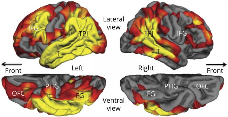
            In the PPA-AD group, participants had a significant yearly decline of 6.21% in object naming scores and 4.25% for a measure of global language performance, with no significant decrease in memory. Those in the DAT-AD group, meanwhile, had a significant yearly decrease of 2.15% for memory and 4.05% for object naming. Structural imaging was also done on the PPA-AD group at their initial visits showing cortical thinning, especially of the language-dominant left hemisphere, extending throughout the language network. Significant thinning of the parahippocampal gyrus was present only on the left side (as shown below, denoted by PHG).

To assess neuropathology, Aβ plaques, neurofibrillary tangles (NFTs), and overall plaque density were quantified. The PPA-AD group showed maximum levels of Aβ and NFTs, though researchers focused on NFTs as their distribution and density more strongly correlates to cognition. Specifically, the PPA-AD group had severe NFT pathology in the neocortex and all medial-temporal lobe structures associated with memory. However, 2 of the 8 who agreed to undergo autopsy were found to be of the “hippocampal-sparing type” where cortical NFT density is higher than in memory-related structures. Interestingly, despite the decrease in hippocampal NFT density and sparing of memory, those 2 participants had severe NFT-induced degeneration of memory regions. Furthermore, bilateral comparisons revealed that the left hemisphere of these 2 participants had more Aβ plaques while other patients had elevated NFTs, suggesting a dichotomy between pathogenesis of hippocampal-sparing type PPA-AD and typical PPA-AD.

            This leaves the question, what induces resilience of memory structures in PPA-AD? Well, in PPA-AD the hippocampal gyrus primarily degenerates on the left hemisphere. Previous lesion studies on this area have shown that episodic memory function only significantly declines when bilateral lesioning or degeneration of hippocampal structures occurs. However, while this study showed decreased NFT aggregation in memory-related structures in PPA-AD compared to cortical areas, previous studies have had varied results indicating that further longitudinal studies of this type are required.

One other possible explanation for memory resilience in PPA-AD involves APOE status. The PPA-AD group had a 14.7% incidence of ε4 alleles, which inhibits neuronal plasticity, while the rest had ε3 alleles, which enhance neuronal plasticity. Interestingly, this frequency of ε4 alleles matches control populations with PPA-AD, while the DAT-AD group had a 42% ε4 frequency with 3 homozygous carriers, almost twice as high as control populations. Thus ε4 presence likely increased vulnerability of memory networks in the DAT-AD group by inhibiting compensatory mechanisms like neuroplasticity, while those with PPA-AD, having reduced ε4 and increased ε3 frequency, may increase resilience to neurodegeneration by allowing for neuronal plasticity.

Facebook
Twitter
Email
Sources:
Sajjadi, S. A., Ash, S., & Cappa, S. Preservation of Memory in Alzheimer’s-Related Primary Progressive Aphasia [Magazine]. Neurology. 2020.
Preib, D., Billette, O. V., Schneider, A., Spotorno, N., & Nestor, P. J. The atrophy pattern in Alzheimer-related PPA is more widespread than that of the frontotemporal lobar degeneration associated variants [Online]. NeuroImage: Clinical. 2019. Available from: https://www.ncbi.nlm.nih.gov/pmc/articles/PMC6734177/
Mesulam, M. M., et al. Memory Resilience in Alzheimer Disease With Primary Progressive Aphasia [Online]. Neurology. 2021. Available from: https://n.neurology.org/content/96/6/e916.long
Primary Progressive Aphasia [Online]. National Aphasia Association. Available from: https://www.aphasia.org/aphasia-resources/primary-progressive-aphasia/

How PWAS Helped Discover 10 New Alzheimer’s Genes

       For quite a while, one of the most common methods of isolating disease-risk-associated genes has been a Genome Wide Association Study (GWAS) which involves genotyping a large number of people and associating various genetic loci, or locations, with the phenotypes, or visible traits, that commonly arise from variance at these loci. However, researchers recently developed an even more informative way of performing these genomic analyses, using it to discover 10 new genes that may modify risk of Alzheimer’s disease (AD)! This new method is very similar to GWAS but adds another layer of data regarding protein function, becoming a Proteome-Wide Association Study (PWAS).

       PWAS expands upon the information provided by GWAS by analyzing the type of mutation present and quantifying the change in production or functionality of the protein produced by that gene in comparison to controls. Next, protein function scores are correlated to the phenotypes of subjects with those genes to confirm the expected effects. In analysis of a binary phenotype (a trait that is either present or absent with no intermediate presentation) a strong correlation is derived when subjects with a disorder have a significantly different functional effect score than controls, confirming PWAS’s prediction that the protein is less (or more) functional in the mutated form.

Figure 1. A diagram depicting the difference in analyses between Genome-Wide Association Studies and Proteome-Wide Association Studies.

       The additional information provided by PWAS allows detection of associations that are not detectable by GWAS. Researchers at Emory University recently took advantage of this, using PWAS to discover 10 new genes associated with AD risk. To begin, they isolated 1,475 genes whose abundance is genetically controlled and analyzed their AD-risk score in a GWAS dataset (71,880 cases, 383,378 controls). Of the 1,475 genes, only 13 were related to AD risk in the GWAS dataset.

       After further analyses with PWAS, including causality tests and adjustments for APOE status, 11 genes remained with evidence for causality of AD. Only 1 of these genes, ACE, had previously been strongly correlated with AD but the other 10 are relatively new discoveries! Those genes include syntaxin 4 and 6, DOC2A, SNX32, ICA1L, cathepsin H, CARHSP1, LACTB, RTFDC1, and PLEKHA1. Although little is known about their relationship to AD pathogenesis, researchers will undoubtedly begin researching how these genes relate to AD risk and development. In time you may even see treatments emerging targeting these genes or proteins.

       Even more interestingly, several of these genes impact molecular pathways that are not widely considered part of the disease model for AD, such as LACTB which is a mitochondrial protein (with mitochondrial dysfunction only getting attention as a possible mechanism of AD development within the last few years) and PLEKHA1 which mediates transmembrane signaling more generally. This suggests that there are as-of-yet undiscovered or under-researched factors that relate to AD risk and pathogenesis, opening the door for new treatments and a more complete understanding of the disease itself.

Facebook
Twitter
Email
Sources:
PWAS x GWAS? Proteome Analysis Nets 10 New Alzheimer’s Genes [Internet]. Alzforum. 2021. Available from: https://www.alzforum.org/news/research-news/pwas-x-gwas-proteome-analysis-nets-10-new-alzheimers-genes
Brandes, N., Linial, N, & Linial, M. PWAS: Proteome-Wide Association Study [Internet]. bioRxiv. 2019. Available from: https://www.biorxiv.org/content/10.1101/812289v1.full