Tau: How Different Isoforms Predict Different Stages of AD Progression

       If you have read our blogs before, you are likely familiar with the two primary biomarkers of Alzheimer’s disease (AD), protein tau which forms neurofibrillary tangles (NFTs) and amyloid beta (Aꞵ) which forms amyloid plaques. Both of these contribute heavily to neuronal dysfunction, degeneration, and eventual memory impairment, but the relationship between them is complicated and has been the subject of research for several years. Evidence suggests that Aꞵ buildup instigates the misfolding of protein tau, eventually inducing NFT formation, however, tau levels better predict cognitive impairment than Aꞵ levels. More recently, researchers have expanded upon this by determining different stages of AD development as predicted by Aꞵ and tau.

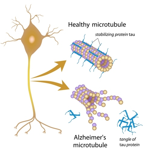
       Before explaining the stages, we need to have some prior knowledge. While we frequently refer to tau as a single protein, this is not necessarily the case. Tau’s full name is microtubule associated protein tau (MAPT), and in its normal form it serves as the rigid scaffolding that helps maintain the shape of axons, the communication bridge between neurons. The diagram below depicts both normal, healthy tau as well as the NFTs that form in cases of AD. It is believed that the presence of toxic Aꞵ proteins induce hyperphosphorylation of tau proteins, changing their structure. This decreases their ability to support microtubules and makes them prone to clumping together, inducing dysfunction both through the tangle of proteins blocking normal cellular functions in the brain and through axonal loss due to their lack of stabilization.

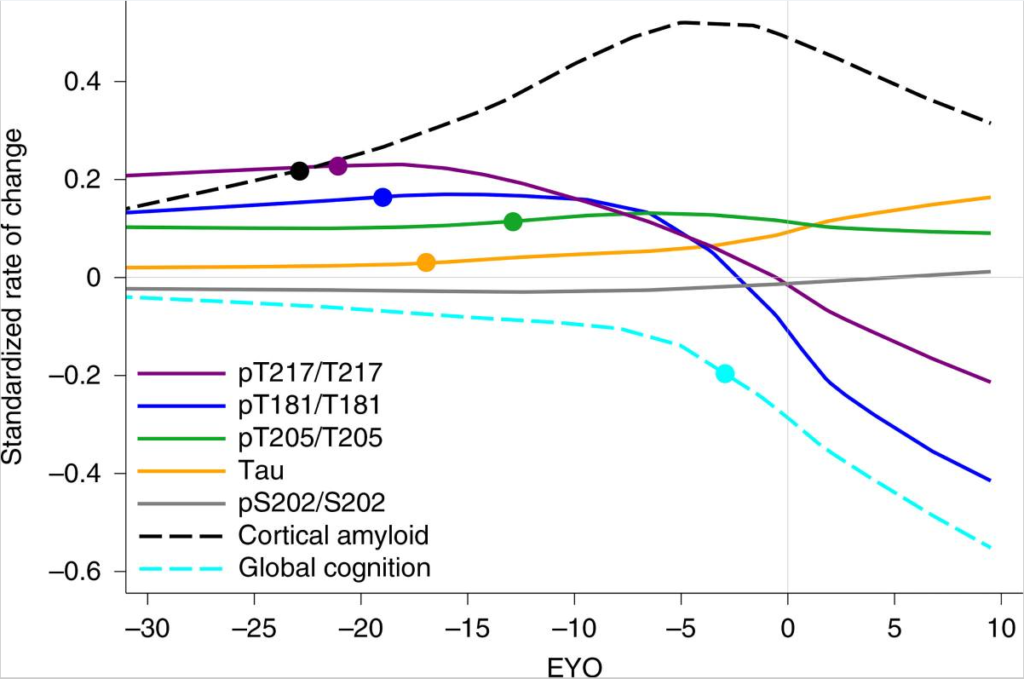
       The specific locations on the protein at which tau can be hyperphosphorylated result in multiple different forms of tau, called p-tau isoforms. The most relevant isoforms to AD are p-tau217, 181, and 205. The presence, or lack thereof, of each type of tau predicts something different and generally correlates to a specific stage of disease progression. For example, an increase of p-tau217 and 181 without presence of NFTs predicts amyloidosis, the buildup of Aꞵ plaques in the brain before symptom onset. A rise of p-tau205 as measured by cerebrospinal fluid (CSF) correlates to waning brain metabolism and shrinking gray matter, the initial stages of degeneration but not yet producing dysfunction. Finally, as total tau levels spike in CSF, NFTs begin to form and cognitive decline begins. Interestingly, once NFT formation begins and global cognition starts to decline, the amount p-tau181 and 217 present in CSF plummets, presumably because these isoforms are being sequestered into the NFTs that are now forming. While this explanation for decrease in CSF p-tau levels is hypothetical, it is supported by the fact that the same phenomenon occurs with amyloid. The figure below from Barthélemy et. al. (2020) exemplifies this sudden change in p-tau and amyloid levels around the estimated year of onset (EYO).

       This information is extremely useful because AD therapies being tested in clinical trials utilize many different mechanisms to fight the disease. Using the different p-tau metrics above, it may be possible to more specifically gauge how far progressed a patient may be and what therapies are most likely to be useful. It is also projected that the increased specificity for placement in trials provided by p-tau measurements, as well as tau PET scans using a new and more accurate tracer, could reduce the sample size needed within clinical trials to find (or disprove) efficacy. Specifically, for trials on preclinical (asymptomatic) AD, using p-tau217 with tau PET scans was hypothesized to reduce required sample size by 43% and by 68% for MCI trials. Using either p-tau217 or tau PET alone would theoretically also result in reduced sample requirements, albeit to a lesser degree, with p-tau217 alone for preclinical AD trials reducing sizes by 31%, and PET alone reducing MCI trial sizes by 47%.

       A decreased sample size with more specific subject selection could provide faster clinical trial outcomes with lessened screening times, and a decrease in the likelihood of a successful drug requiring additional data before coming to market. Should these staging procedures become a widespread method of pre-screening, patients are more likely to be placed into a clinical trial that will help them based upon their specific disease staging, whether that be clearing tau tangles, preventing tau aggregation, or clearing amyloid proteins before they even initiate the hyperphosphorylation of tau.

Facebook
Twitter
Email
Sources:
Different CSF Phospho-Taus Match Distinct Changes in Brain Pathology. Alzforum [Internet]. 2020. Available from: https://www.alzforum.org/news/research-news/different-csf-phospho-taus-match-distinct-changes-brain-pathology
In Preclinical Alzheimer’s, p-tau217 in Blood Best Predicts Tangles. Alzforum [Internet]. 2021. Available from: https://www.alzforum.org/news/research-news/preclinical-alzheimers-p-tau217-blood-best-predicts-tangles
Barthélemy et. al. A soluble phosphorylated tau signature links tau, amyloid and the evolution of stages of dominantly inherited Alzheimer’s disease. Nature Medicine [Internet]. 2020. Available from: https://www.ncbi.nlm.nih.gov/pmc/articles/PMC7309367/

Insulin Resistance and Alzheimer’s: A Two Way Street (and How GLP-1 Receptor Agonists May Help Cross It)

       Previously, we described the relationship between insulin resistance and AD, and treatments pertaining to such (https://www.centerforcognitivehealth.com/insulin-and-ad/). However, the overarching principle of how insulin signaling ties into development of neurodegenerative conditions is only loosely understood, indicating the need for further research.

       Insulin, produced by the pancreas, signals to maintain glucose homeostasis and cell growth/survival by binding to insulin receptors (IRs). Insulin resistance is caused by a downregulation of these IRs, which in turn instigates an overproduction of insulin (hyperinsulinemia) to try to overcome the limited signaling. IRs are present in large quantities in the brain, especially in the hippocampus, a prominent structure for memory. During hyperinsulinemia episodes our bodies downregulate the transporters that allow insulin into the brain, possibly increasing cell death, decreasing cell growth, and impairing memory. Diseases, such as AD and Parkinson’s disease (PD), are twice as likely to develop in individuals with diabetes, supporting this relationship.

       While diabetes increases the risk of AD, AD also increases the risk of developing type II diabetes mellitus (T2DM). Research into AD’s role in causing T2DM showed that toxic amyloid-ꞵ (Aꞵ) oligomers in the AD brain interact with hippocampal tissues to reduce the number of IRs present, and is predictive of insulin resistance outside the brain, eventually inducing T2DM. Furthermore, inflammation is strongly tied to the development of both T2DM and AD, possibly explained by the fact that insulin resistance increases circulating inflammatory cytokines.

       It appears that treating peripheral insulin resistance has both a direct and indirect impact on risk/prevention of AD on top of the obvious impact on diabetes/insulin resistance. Clinical trials aimed at treating AD have taken notice. For instance, we currently have a trial utilizing semaglutide, a medication already approved as an antidiabetic treatment, to attempt to stop/slow progression of AD in individuals with Mild Cognitive Impairment or Early AD. A hormone called GLP-1 has also been implicated in playing a role in both diabetes and AD. GLP-1 is similar to insulin with a strong role in glucose homeostasis but is quickly degraded under normal circumstances. Semaglutide, a GLP-1 receptor agonist (RA), simulates the effects of GLP-1 while avoiding quick degradation, creating lasting impacts on glucose regulation without being impacted by insulin resistance.

       Before semaglutide, several other molecules were tested for this purpose. The first GLP-1 RA, exendin-4, improved cognition and reduced Aꞵ presence in the brains of both AD mice and wild-type mice. The next major GLP-1 RA, liraglutide, produced longer lasting effects than exendin-4 and was shown to prevent Aꞵ neurotoxicity and reduce Aꞵ plaques in the hippocampus and cortex, reduce cell death, alleviate brain insulin resistance, and improve memory in the same mouse model exendin-4 was tested on. It also lowered levels of phosphorylated Tau, the other major protein implicated in AD progression. When administered before significant plaque burden was present and memory impairment began, liraglutide slowed disease progression in AD mouse models. Yet another marketed diabetes drug, lixisenatide, enhances long-term potentiation, and lowers Aꞵ plaque load, microglial activation, and neurofibrillary tangles. Despite these other treatments, semaglutide shows the greatest effectiveness compared to other GLP-1 RAs with regards to glycemic regulation. Given how intertwined insulin resistance and neurodegeneration seem to be, greater efficacy in one instance may offer benefit in the other. Furthermore, semaglutide is already approved and marketed for treatment of diabetes so it’s safety and tolerability are well studied.

       With all this therapeutic potential surrounding semaglutide and GLP-1 RAs, if you or someone you know between the ages of 55 and 85 is experiencing Mild Cognitive Impairment (MCI) or mild Alzheimer’s dementia they may be eligible to screen for the trial! Feel free to contact our office or inquire about potential involvement on our website.

Facebook
Twitter
Email
Sources:
Batista, A.F., Bodart-Santos, V., De Felice, F. G., & Ferreira, S.T. Neuroprotective Actions of Glucagon-Like Peptide-1 (GLP-1) Analogues in Alzheimer’s and Parkinson’s Diseases [Internet]. CNS Drugs. 2018. Available from: https://www.researchgate.net/publication/329373799_Neuroprotective_Actions_of_Glucagon-Like_Peptide-1_GLP-1_Analogues_in_Alzheimer’s_and_Parkinson’s_Diseases
Insulin and Alzheimer’s Disease [Blog]. 2020. Available from: https://www.centerforcognitivehealth.com/insulin-and-ad/
Alsugair, H.A., Alshugair, I.F., Alharbi, T.J., Bin Rsheed, A.M., Tourkmani, A.M., Al-Madani, W. Weekly Semaglutide vs. Liraglutide Efficacy Profile: A Network Meta-Analysis [Internet]. Healthcare. 2021. Available from: https://pubmed.ncbi.nlm.nih.gov/34574899/