What Are Coarse-Grained Plaques?

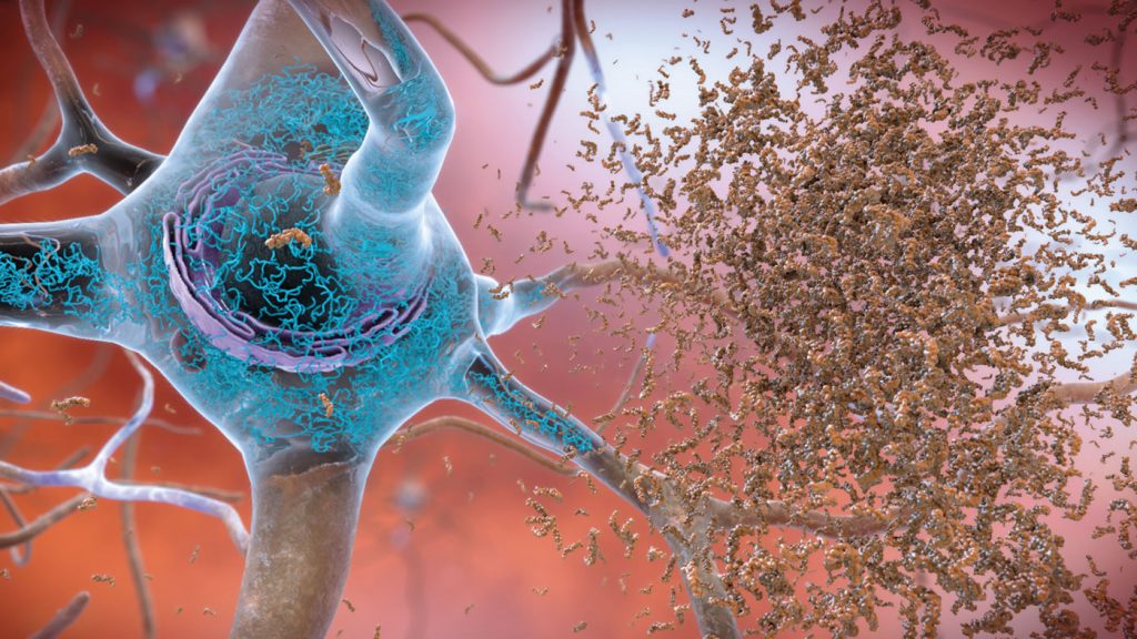
       Alzheimer’s disease (AD) was identified in 1901 by Alois Alzheimer but despite being known for over a century, researchers are discovering new things about the disease today. For example, our regular readers should be familiar with the term “amyloid plaques” which are likely the most well researched biomarkers of AD. There are also sub-types of amyloid plaques and a group of pathologists recently observed what they believe to be a unique sub-type of plaque termed “coarse-grained plaques”. This week we will discuss these new plaques and their significance to AD research.

       Coarse-grained plaques form a core of amyloid-ꞵ with 42 residues (Aꞵ42) surrounded by a shell of Aꞵ40, and are packed with a protein called norrin, a marker of blood-vessel damage. These plaques tend to localize near blood vessels but cannot cross the blood-brain barrier (BBB). 84% of the analyzed coarse-grained plaques had direct contact with vasculature, suggesting that they may induce cerebrovascular dysfunction rather than neurological dysfunction directly.

       They are more common in people with two copies of the ApoE4 allele or those with early onset AD. In this study out of 28 non-ApoE4 carriers, 15 had sparse/frequent coarse-grained plaques, compared to 25 out of 33 for heterozygous ApoE4 carriers. All 11 homozygous carriers had moderate/frequent deposition of the coarse-grained plaques. This suggests interaction between the ApoE gene and coarse-grained plaques. In fact, there may even be a separate ApoE4-induced AD sub-type in homozygous carriers.

       Researchers used laser scanning microscopy to visualize the plaques and saw that out of 74 brains, 38 had early onset AD, 21 had late onset, and 15 were never diagnosed with dementia though amyloid positive. This diverse grouping shows that, while coarse-grained plaques might induce early AD in many cases, it does not guarantee a specific pathological presentation. Staining techniques and complement testing were utilized to discover more about the plaques on a molecular level.

       The stained coarse-grained plaques showed amyloid precursor protein (APP) and prion protein suggesting they may damage nearby neurons. The complement testing also revealed that these plaques have markers for extreme neuroinflammation and presence of astrocytes and microglia. In fact, microglia and astrocytes cover these plaques in a particular pattern which seems to further differentiate this type of plaque from those which were known previously, confirming the unique nature of these new plaques.

       In summary, these specific plaques are only just beginning to be researched but already it seems there is a close relationship between the presence of coarse-grained plaques, the ApoE4 allele, cerebrovasculature, and AD pathology. With more studies and larger sample sizes, research of this topic may lead to innovation in the diagnosis and differing treatment options for subtypes of AD.

Facebook
Twitter
Email
Source:
Introducing the Coarse-Grained Plaque – A New Type of Amyloid [Internet].Alzforum. 2020. Available from: https://www.alzforum.org/news/research-news/introducing-coarse-grained-plaque-new-type-amyloid

Cognitive Resilience: Developmental Vs. Genetic Factors

       A recent discovery of a genetically unique family has shown that developmental disorders may predispose or change the presentation of Alzheimer’s disease (AD) dementia. The family studied consisted of 10 siblings, 8 of whom presented with developmental language problems and 1 with a sub-syndrome of frontotemporal dementia known as logopenic variant primary progressive aphasia (lvPPA). All members had average total brain volumes but decreased left cortical volume, and reduced functional connectivity between the left superior temporal cortex (responsible for auditory processing) and language areas in both hemispheres resulting in the language dysfunction.

       The researchers hypothesized that developmental delays in language and changes in brain morphology may induce AD symptomology effectively matching that of lvPPA. Due to the pre-existing changes and vulnerability of the language network, these networks are first to dysfunction. In those without developmental language delays, AD selectively affects executive functioning first. This contrasting pathology shows that pre-existing conditions and morphological changes in the brain might interact to change or accelerate the development of other disorders.

       If pre-existing dysfunctional networks contribute to the development of AD, it’s possible hyper-functional networks may be neuroprotective. To assess this theory elderly participants received biennial amyloid PET scans and underwent yearly cognitive testing for 14 years. The presence of the APOE-2 gene, lower pulse pressure, and higher baseline scores on cognitive tests appeared to be neuroprotective. Engagement in paid work and increased life satisfaction also predicted resilience to cognitive decline, but to a lesser degree. Furthermore, in amyloid-positive participants, never having smoked also predicted cognitive resilience.

       While the APOE-2 genotype is neuroprotective via reduced likelihood of amyloid build-up, the relationship between pulse pressure, cognitive testing scores, and AD are in need of further study. In those who are amyloid positive, higher baseline cognitive scores and never having smoked predicted increased cognitive resilience. This suggests that networks with greater functional connectivity (in comparison to the family with language deficits who had decreased connectivity) are indeed able to remain cognitively normal for longer. Additionally, cerebral vasculature appears to play an important role in cognitive resilience, with those with lower pulse pressure remaining cognitively normal for longer. On top of this, smoking increases blood pressure further supporting the concept that those who never smoked were also more cognitively resilient.

       Genetics are only one factor in the multifaceted disorder that is AD. Presence of amyloid does not guarantee AD pathology, AD pathology may present differently in a brain with morphological and functional changes induced by other genes, and non-genetic factors such as smoking and lifestyle also play a role. Researchers are now tasked with finding the intersection of genetics, lifestyle, and comorbid disorders to determine how these things influence AD and from there, how to counteract cognitive decline or even prevent it in the first place!

Facebook
Twitter
Email
Sources:
Hillis, A. E. & Kolundžić, Z. Developmental and degenerative deficiencies in the language network [Internet]. Neurology. 2020. Available from: https://n.neurology.org/content/95/7/281
Snitz, B. E. et al. Predicting resistance to amyloid-beta deposition and cognitive resilience in the oldest-old [Internet]. Neurology. 2020. Available from: https://n.neurology.org/content/95/8/e984

Insulin and Alzheimer’s Disease

Insulin

        Insulin is typically associated with regulating blood glucose levels and diabetes, but it also serves as a crucial signaling molecule throughout the body, including the central nervous system (CNS). In fact, there is evidence that insulin may play a role in the development of Alzheimer’s disease (AD) and Mild Cognitive Impairment (MCI). Dysregulation of insulin signaling negatively impacts cognition and increases deposition of amyloid plaques and tau tangles. Luckily, there are also mechanisms that can upregulate insulin sensitivity and these treatments have potential to reduce or alleviate AD symptoms.

       Insulin is a hormone released primarily by the pancreas. It can cross the blood-brain barrier (BBB) to have CNS effects. However, if one develops insulin resistance, which is correlated with AD, normal tissues fail to sufficiently respond to the presence of insulin. In these cases, insulin dysregulation induces hyperglycemia, too much sugar in the blood, which leads to glucose neurotoxicity, reduced cerebral blood flow, and accumulation of toxic byproducts in the brain, all of which can lead to cognitive impairment.

       There are several ways to test insulin resistance but they all follow a basic method of administering insulin and monitoring the response of the target tissue, whether peripheral or in the CNS. There is a close bi-directional relationship between insulin functioning in the body and the brain. For patients with AD there is a trend towards insulin insensitivity and hyperinsulinemia (too much insulin in the blood). Excess insulin downregulates the receptors that move insulin through the BBB into the CNS, meaning less insulin in the brain.

       A reduction in CNS insulin is significant because insulin in the brain protects against amyloid-beta synaptotoxicity and promotes clearance of plaques. AD patients with peripheral insulin resistance have increased amyloid deposition compared to healthy controls. Tests of peripheral insulin resistance successfully predict amyloid deposition in the brain 15 years later, as confirmed by an amyloid PET scan. Furthermore, patients with altered insulin signaling from diabetes have increased tau levels in cerebrospinal fluid. A final additive risk factor is that excess insulin acts as a vasoconstrictor limiting blood flow to the brain and decreasing amyloid and tau clearance. Dysfunctional insulin signaling may be a risk factor for AD.

       Understanding how insulin resistance impacts AD risk has improved our range of potential treatments. Intranasal insulin administration allows it to bypass the BBB and enter the CNS, reducing AD pathology and improving memory in rats after long-term administration. In humans, twenty-one days of treatment enhanced episodic memory. However, clinical trials testing intranasal insulin show mixed results indicating the need for further research. One can also increase insulin sensitivity with “insulin sensitizers”, such as Metformin, but evidence for these treatments in AD is limited. In mice and primates a GLP-1 agonist, which stimulates insulin production and regulates glucostasis, called liraglutide preserved memory and increased hippocampal neuronal density. Liraglutide is currently in a Phase II clinical trial for use in humans with AD.

       Although these potential treatments are promising there are proven ways to enhance insulin sensitivity and reduce AD risk right now and without a prescription. Consuming a diet of primarily polyunsaturated fatty acids, nuts, and plant-based foods correlates with increased insulin sensitivity and decreased risk of AD-related cognitive decline compared to diets containing higher saturated fats, animal proteins, and refined sugars. Additionally, regular exercise is a powerful modulator of insulin sensitivity and has also been shown to reduce risk of AD. In the meantime, while we wait for the aforementioned therapies to be approved, a lifestyle and diet change is not only protective against AD, but can also improve your overall general health.

Facebook
Twitter
Email
Sources:
Kellar, D. & Craft, S. Brain insulin resistance in Alzheimer’s disease and related disorders: mechanisms and therapeutic approaches [Internet]. The Lancet Neurology. 2020. Available from: https://www.sciencedirect.com/science/article/abs/pii/S1474442220302313